Structural basis for compound C inhibition of the human AMP-activated protein kinase alpha 2 subunit kinase domain
Handa, N., Takagi, T., Saijo, S., Kishishita, S., Takaya, D., Toyama, M., Terada, T., Shirouzu, M., Suzuki, A., Lee, S., Yamauchi, T., Okada-Iwabu, M., Iwabu, M., Kadowaki, T., Minokoshi, Y., Yokoyama, S.(2011) Acta Crystallogr D Biol Crystallogr 67: 480-487
- PubMed: 21543851
- DOI: https://doi.org/10.1107/S0907444911010201
- Primary Citation of Related Structures:
2YZA, 3AQV - PubMed Abstract:
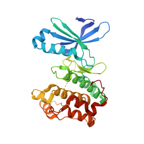
AMP-activated protein kinase (AMPK) is a serine/threonine kinase that functions as a sensor to maintain energy balance at both the cellular and the whole-body levels and is therefore a potential target for drug design against metabolic syndrome, obesity and type 2 diabetes. Here, the crystal structure of the phosphorylated-state mimic T172D mutant kinase domain from the human AMPK α2 subunit is reported in the apo form and in complex with a selective inhibitor, compound C. The AMPK α2 kinase domain exhibits a typical bilobal kinase fold and exists as a monomer in the crystal. Like the wild-type apo form, the T172D mutant apo form adopts the autoinhibited structure of the `DFG-out' conformation, with the Phe residue of the DFG motif anchored within the putative ATP-binding pocket. Compound C binding dramatically alters the conformation of the activation loop, which adopts an intermediate conformation between DFG-out and DFG-in. This induced fit forms a compound-C binding pocket composed of the N-lobe, the C-lobe and the hinge of the kinase domain. The pocket partially overlaps with the putative ATP-binding pocket. These three-dimensional structures will be useful to guide drug discovery.
Organizational Affiliation:
RIKEN Systems and Structural Biology Center, 1-7-22 Suehiro-cho, Tsurumi, Yokohama 230-0045, Japan.