2XFO

tranylcypromine-inhibited human monoamine oxidase B Ile199Ala mutant in complex with 2-(2-benzofuranyl)-2-imidazoline


Experimental Data Snapshot

  • Method: X-RAY DIFFRACTION
  • Resolution: 2.10 Å
  • R-Value Free: 0.243 
  • R-Value Work: 0.189 
  • R-Value Observed: 0.190 

wwPDB Validation   3D Report Full Report


Ligand Structure Quality Assessment 


This is version 1.6 of the entry. See complete history


Literature

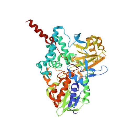
Potentiation of ligand binding through cooperative effects in monoamine oxidase B.

Bonivento, D.Milczek, E.M.McDonald, G.R.Binda, C.Holt, A.Edmondson, D.E.Mattevi, A.

(2010) J Biol Chem 285: 36849-36856

  • DOI: https://doi.org/10.1074/jbc.M110.169482
  • Primary Citation of Related Structures:  
    2XCG, 2XFN, 2XFO, 2XFP, 2XFQ, 2XFU

  • PubMed Abstract: 

    Crystallographic and biochemical studies have been employed to identify the binding site and mechanism for potentiation of imidazoline binding in human monoamine oxidase B (MAO B). 2-(2-Benzofuranyl)-2-imidazoline (2-BFI) inhibits recombinant human MAO B with a K(i) of 8.3 ± 0.6 μM, whereas tranylcypromine-inhibited MAO B binds 2-BFI with a K(d) of 9 ± 2 nM, representing an increase in binding energy Δ(ΔG) of -3.9 kcal/mol. Crystal structures show the imidazoline ligand bound in a site that is distinct from the substrate-binding cavity. Contributions to account for the increase in binding affinity upon tranylcypromine inhibition include a conformational change in the side chain of Gln(206) and a "closed conformation" of the side chain of Ile(199), forming a hydrophobic "sandwich" with the side chain of Ile(316) on each face of the benzofuran ring of 2-BFI. Data with the I199A mutant of human MAO B and failure to observe a similar binding potentiation with rat MAO B, where Ile(316) is replaced with a Val residue, support an allosteric mechanism where the increased binding affinity of 2-BFI results from a cooperative increase in H-bond strength through formation of a more hydrophobic milieu. These insights should prove valuable in the design of high affinity and specific reversible MAO B inhibitors.


  • Organizational Affiliation

    Department of Genetics and Microbiology, University of Pavia, I-27100 Pavia PV, Italy.


Macromolecules
Find similar proteins by:  (by identity cutoff)  |  3D Structure
Entity ID: 1
MoleculeChains Sequence LengthOrganismDetailsImage
Amine oxidase [flavin-containing] B
A, B
520Homo sapiensMutation(s): 1 
Gene Names: MAOB
EC: 1.4.3.4
Membrane Entity: Yes 
UniProt & NIH Common Fund Data Resources
Find proteins for P27338 (Homo sapiens)
Explore P27338 
Go to UniProtKB:  P27338
PHAROS:  P27338
GTEx:  ENSG00000069535 
Entity Groups  
Sequence Clusters30% Identity50% Identity70% Identity90% Identity95% Identity100% Identity
UniProt GroupP27338
Sequence Annotations
Expand
  • Reference Sequence
Small Molecules
Ligands 4 Unique
IDChains Name / Formula / InChI Key2D Diagram3D Interactions
FA8
Query on FA8

Download Ideal Coordinates CCD File 
C [auth A][[(2R,3S,4S)-5-[(4AS)-7,8-DIMETHYL-2,4-DIOXO-4A,5-DIHYDROBENZO[G]PTERIDIN-10-YL]-2,3,4-TRIHYDROXY-PENTOXY]-HYDROXY-PHOSPHORYL] [(2R,3S,4R,5R)-5-(6-AMINOPURIN-9-YL)-3,4-DIHYDROXY-OXOLAN-2-YL]METHYL HYDROGEN PHOSPHATE
C27 H35 N9 O15 P2
SDBHOOIZTSVSKT-LDVRFEIFSA-N
FAD
Query on FAD

Download Ideal Coordinates CCD File 
F [auth B]FLAVIN-ADENINE DINUCLEOTIDE
C27 H33 N9 O15 P2
VWWQXMAJTJZDQX-UYBVJOGSSA-N
XCG
Query on XCG

Download Ideal Coordinates CCD File 
E [auth A],
H [auth B]
2-(2-BENZOFURANYL)-2-IMIDAZOLINE
C11 H8 N2 O
VBFQIUOSXHUWCT-UHFFFAOYSA-N
3PL
Query on 3PL

Download Ideal Coordinates CCD File 
D [auth A],
G [auth B]
3-PHENYLPROPANAL
C9 H10 O
YGCZTXZTJXYWCO-UHFFFAOYSA-N
Binding Affinity Annotations 
IDSourceBinding Affinity
XCG Binding MOAD:  2XFO Ki: 5.80e+4 (nM) from 1 assay(s)
Experimental Data & Validation

Experimental Data

  • Method: X-RAY DIFFRACTION
  • Resolution: 2.10 Å
  • R-Value Free: 0.243 
  • R-Value Work: 0.189 
  • R-Value Observed: 0.190 
  • Space Group: C 2 2 2
Unit Cell:
Length ( Å )Angle ( ˚ )
a = 130.546α = 90
b = 221.863β = 90
c = 86.033γ = 90
Software Package:
Software NamePurpose
REFMACrefinement
MOSFLMdata reduction
SCALAdata scaling
REFMACphasing

Structure Validation

View Full Validation Report


Ligand Structure Quality Assessment 


Entry History 

Deposition Data

Revision History  (Full details and data files)

  • Version 1.0: 2010-10-06
    Type: Initial release
  • Version 1.1: 2011-05-08
    Changes: Version format compliance
  • Version 1.2: 2011-07-13
    Changes: Version format compliance
  • Version 1.3: 2017-12-20
    Changes: Database references
  • Version 1.4: 2018-05-16
    Changes: Data collection
  • Version 1.5: 2018-07-25
    Changes: Data collection, Database references, Source and taxonomy, Structure summary
  • Version 1.6: 2023-12-20
    Changes: Data collection, Database references, Derived calculations, Other, Refinement description