2WTY

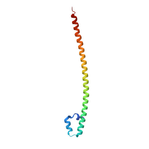
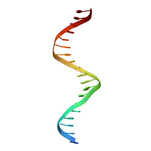
Crystal structure of the homodimeric MafB in complex with the T-MARE binding site


Experimental Data Snapshot

  • Method: X-RAY DIFFRACTION
  • Resolution: 2.90 Å
  • R-Value Free: 0.275 
  • R-Value Work: 0.235 
  • R-Value Observed: 0.237 

wwPDB Validation   3D Report Full Report


This is version 1.2 of the entry. See complete history


Literature

Design of a bZIP Transcription Factor with Homo/Heterodimer-Induced DNA-Binding Preference.

Pogenberg, V.Consani Textor, L.Vanhille, L.Holton, S.J.Sieweke, M.H.Wilmanns, M.

(2014) Structure 22: 466

  • DOI: https://doi.org/10.1016/j.str.2013.12.017
  • Primary Citation of Related Structures:  
    2WT7, 2WTY

  • PubMed Abstract: 

    The ability of basic leucine zipper transcription factors for homo- or heterodimerization provides a paradigm for combinatorial control of eukaryotic gene expression. It has been unclear, however, how facultative dimerization results in alternative DNA-binding repertoires on distinct regulatory elements. To unravel the molecular basis of such coupled preferences, we determined two high-resolution structures of the transcription factor MafB as a homodimer and as a heterodimer with c-Fos bound to variants of the Maf-recognition element. The structures revealed several unexpected and dimer-specific coiled-coil-heptad interactions. Based on these findings, we have engineered two MafB mutants with opposite dimerization preferences. One of them showed a strong preference for MafB/c-Fos heterodimerization and enabled selection of heterodimer-favoring over homodimer-specific Maf-recognition element variants. Our data provide a concept for transcription factor design to selectively activate dimer-specific pathways and binding repertoires.


  • Organizational Affiliation

    EMBL Hamburg c/o DESY, Notkestraße 85, 22603 Hamburg, Germany.


Macromolecules

Find similar proteins by:  (by identity cutoff)  |  3D Structure
Entity ID: 1
MoleculeChains Sequence LengthOrganismDetailsImage
TRANSCRIPTION FACTOR MAFB
A, B
97Mus musculusMutation(s): 2 
UniProt
Find proteins for P54841 (Mus musculus)
Explore P54841 
Go to UniProtKB:  P54841
Entity Groups  
Sequence Clusters30% Identity50% Identity70% Identity90% Identity95% Identity100% Identity
UniProt GroupP54841
Sequence Annotations
Expand
  • Reference Sequence

Find similar nucleic acids by:  Sequence   |   3D Structure  

Entity ID: 2
MoleculeChains LengthOrganismImage
DNA (5'-D(*TP*AP*AP*TP*TP*GP*CP*TP*GP*AP*CP*TP*CP*AP *GP*CP*AP*AP*AP*T)-3')20N/A
Sequence Annotations
Expand
  • Reference Sequence

Find similar nucleic acids by:  Sequence   |   3D Structure  

Entity ID: 3
MoleculeChains LengthOrganismImage
DNA (5'-D(*TP*AP*TP*TP*TP*GP*CP*TP*GP*AP*GP*TP*CP*AP *GP*CP*AP*AP*TP*T)-3')20N/A
Sequence Annotations
Expand
  • Reference Sequence
Small Molecules
Ligands 1 Unique
IDChains Name / Formula / InChI Key2D Diagram3D Interactions
MG
Query on MG

Download Ideal Coordinates CCD File 
E [auth D]MAGNESIUM ION
Mg
JLVVSXFLKOJNIY-UHFFFAOYSA-N
Experimental Data & Validation

Experimental Data

  • Method: X-RAY DIFFRACTION
  • Resolution: 2.90 Å
  • R-Value Free: 0.275 
  • R-Value Work: 0.235 
  • R-Value Observed: 0.237 
  • Space Group: P 2 21 21
Unit Cell:
Length ( Å )Angle ( ˚ )
a = 41.15α = 90
b = 49.82β = 90
c = 170.37γ = 90
Software Package:
Software NamePurpose
REFMACrefinement

Structure Validation

View Full Validation Report


Entry History 

Deposition Data

Revision History  (Full details and data files)

  • Version 1.0: 2010-12-08
    Type: Initial release
  • Version 1.1: 2014-02-26
    Changes: Database references, Source and taxonomy, Version format compliance
  • Version 1.2: 2014-03-19
    Changes: Database references