2WPU

Chaperoned ruthenium metallodrugs that recognize telomeric DNA


Experimental Data Snapshot

  • Method: X-RAY DIFFRACTION
  • Resolution: 1.92 Å
  • R-Value Free: 0.198 
  • R-Value Work: 0.172 
  • R-Value Observed: 0.173 

wwPDB Validation   3D Report Full Report


Ligand Structure Quality Assessment 


This is version 1.5 of the entry. See complete history


Literature

Chemo-Genetic Optimization of DNA Recognition by Metallodrugs Using a Presenter-Protein Strategy.

Zimbron, J.M.Sardo, A.Heinisch, T.Wohlschlager, T.Gradinaru, J.Massa, C.Schirmer, T.Creus, M.Ward, T.R.

(2010) Chemistry 16: 12883

  • DOI: https://doi.org/10.1002/chem.201001573
  • Primary Citation of Related Structures:  
    2WPU

  • PubMed Abstract: 

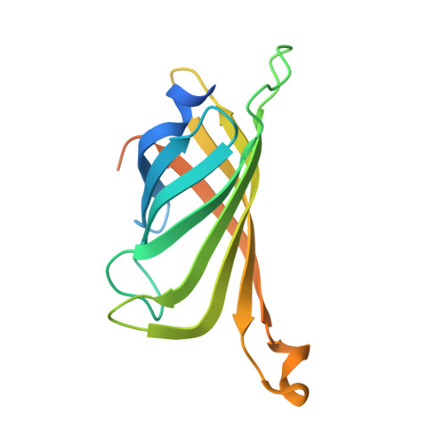
    The mode of action of precious metal anticancer metallodrugs is generally believed to involve DNA as a target. However, the poor specificity of such drugs often requires high doses and leads to undesirable side-effects. With the aim of improving the specificity of a ruthenium piano-stool complex towards DNA, we employed a presenter protein strategy based on the biotin-avidin technology. Guided by the X-ray structure of the assembly of streptavidin and a biotinylated piano-stool, we explored the formation of metallodrug-mediated ternary complexes with the presenter protein and DNA. The assemblies bound more strongly to telomere G-quadruplexes than to double-stranded DNA; chemo-genetic modifications (varying the complex or mutating the protein) modulated binding to these targets. We suggest that rational targeting of small molecules by presenter proteins could be exploited to bind metallodrugs to preferred macromolecular targets.


  • Organizational Affiliation

    University of Basel, Department of Chemistry, Spitalstrasse 51, 4056 Basel, Switzerland.


Macromolecules
Find similar proteins by:  (by identity cutoff)  |  3D Structure
Entity ID: 1
MoleculeChains Sequence LengthOrganismDetailsImage
STREPTAVIDIN159Streptomyces avidiniiMutation(s): 0 
UniProt
Find proteins for P22629 (Streptomyces avidinii)
Explore P22629 
Go to UniProtKB:  P22629
Entity Groups  
Sequence Clusters30% Identity50% Identity70% Identity90% Identity95% Identity100% Identity
UniProt GroupP22629
Sequence Annotations
Expand
  • Reference Sequence
Small Molecules
Ligands 3 Unique
IDChains Name / Formula / InChI Key2D Diagram3D Interactions
KYT
Query on KYT

Download Ideal Coordinates CCD File 
B [auth A](3AS,4S,6AR)-4-(5-((3R,4R)-3,4-DIAMINOPYRROLIDIN-1-YL)-5-OXOPENTYL)TETRAHYDRO-1H-THIENO[3,4-D]IMIDAZOL-2(3H)-ONE-P-CYMENE-CHLORO-RUTHENIUM(III)
C24 H39 Cl N5 O2 Ru S
OJIBJWGHGYGDGX-SNKMZJELSA-M
SO4
Query on SO4

Download Ideal Coordinates CCD File 
C [auth A]SULFATE ION
O4 S
QAOWNCQODCNURD-UHFFFAOYSA-L
GOL
Query on GOL

Download Ideal Coordinates CCD File 
D [auth A]GLYCEROL
C3 H8 O3
PEDCQBHIVMGVHV-UHFFFAOYSA-N
Binding Affinity Annotations 
IDSourceBinding Affinity
KYT Binding MOAD:  2WPU Kd: 60 (nM) from 1 assay(s)
Experimental Data & Validation

Experimental Data

  • Method: X-RAY DIFFRACTION
  • Resolution: 1.92 Å
  • R-Value Free: 0.198 
  • R-Value Work: 0.172 
  • R-Value Observed: 0.173 
  • Space Group: I 41 2 2
Unit Cell:
Length ( Å )Angle ( ˚ )
a = 57.68α = 90
b = 57.68β = 90
c = 183.694γ = 90
Software Package:
Software NamePurpose
REFMACrefinement
MOSFLMdata reduction
SCALAdata scaling
MOLREPphasing

Structure Validation

View Full Validation Report


Ligand Structure Quality Assessment 


Entry History 

Deposition Data

Revision History  (Full details and data files)

  • Version 1.0: 2010-10-13
    Type: Initial release
  • Version 1.1: 2011-05-08
    Changes: Version format compliance
  • Version 1.2: 2011-07-13
    Changes: Version format compliance
  • Version 1.3: 2019-01-30
    Changes: Data collection, Experimental preparation, Other
  • Version 1.4: 2019-02-06
    Changes: Data collection, Experimental preparation
  • Version 1.5: 2023-12-20
    Changes: Data collection, Database references, Derived calculations, Other, Refinement description