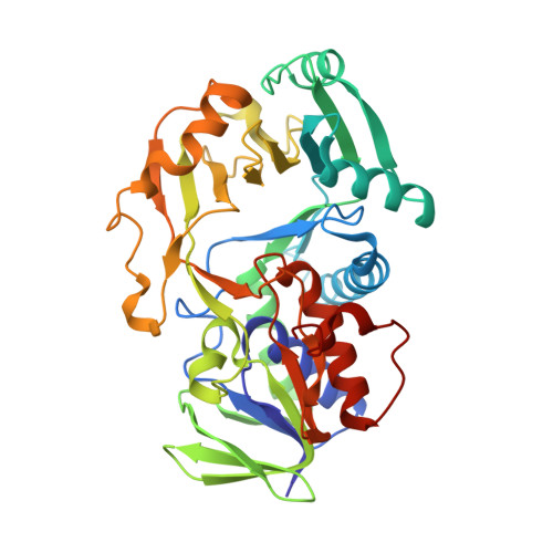
The X-Ray Structure of N-Methyltryptophan Oxidase Reveals the Structural Determinants of Substrate Specificity.
Ilari, A., Bonamore, A., Franceschini, S., Fiorillo, A., Boffi, A., Colotti, G.(2008) Proteins 71: 2065
- PubMed: 18186483
- DOI: https://doi.org/10.1002/prot.21898
- Primary Citation of Related Structures:
2UZZ - PubMed Abstract:
The X-ray structure of monomeric N-methyltryptophan oxidase from Escherichia coli (MTOX) has been solved at 3.2 A resolution by molecular replacement methods using Bacillus sp. sarcosine oxidase structure (MSOX, 43% sequence identity) as search model. The analysis of the substrate binding site highlights the structural determinants that favour the accommodation of the bulky N-methyltryptophan residue in MTOX. In fact, although the nature and geometry of the catalytic residues within the first contact shell of the FAD moiety appear to be virtually superposable in MTOX and MSOX, the presence of a Thr residue in position 239 in MTOX (Met245 in MSOX) located at the entrance of the active site appears to play a key role for the recognition of the amino acid substrate side chain. Accordingly, a 15 fold increase in k(cat) and 100 fold decrease in K(m) for sarcosine as substrate has been achieved in MTOX upon T239M mutation, with a concomitant three-fold decrease in activity towards N-methyltryptophan. These data provide clear evidence for the presence of a catalytic core, common to the members of the methylaminoacid oxidase subfamily, and of a side chain recognition pocket, located at the entrance of the active site, that can be adjusted to host diverse aminoacids in the different enzyme species. The site involved in the covalent attachment of flavin has also been addressed by screening degenerate mutants in the relevant positions around Cys308-FAD linkage. Lys341 appears to be the key residue involved in flavin incorporation and covalent linkage.
Organizational Affiliation:
Institute of Molecular Biology and Pathology, C.N.R., Rome, Italy.