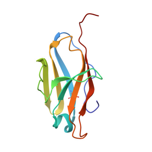
Structure of a novel Bence-Jones protein (Rhe) fragment at 1.6 A resolution.
Furey Jr., W., Wang, B.C., Yoo, C.S., Sax, M.(1983) J Mol Biol 167: 661-692
- PubMed: 6876161
- DOI: https://doi.org/10.1016/s0022-2836(83)80104-1
- Primary Citation of Related Structures:
2RHE - PubMed Abstract:
The crystal structure of Rhe, a lambda-type Bence-Jones protein fragment, has been solved and refined to a resolution of 1.6 A. A model fragment consisting of the complete variable domain and the first three residues of the constant domain yields a crystallographic residual RF value of 0.149. The protein exists as a dimer both in solution and in the crystals. Although the "immunoglobulin fold" is generally preserved in the structure, there are significant differences in both the monomer conformation and in the mode of association of monomers into dimers, when compared to other known Bence-Jones proteins or Fab fragments. The variations in conformation within monomers are particularly significant as they involve non-hypervariable residues, which previously were believed to be part of a "structurally invariant" framework common to all immunoglobulin variable domains. The novel mode of dimerization is equally important, as it can result in combining site shapes and sizes unobtainable with the conventional mode of dimerization. A comparison of the structure with other variable domain dimers reveals further that the variations within monomers and between domains in the dimer are coupled. Some possible functional implications revealed by this coupling are greater variability, induced fitting of the combining site to better accommodate antigenic determinants, and a mechanism for relaying binding information from one end of the variable domain dimer to the other. In addition to providing the most accurate atomic parameters for an immunoglobulin domain yet obtained, the high resolution and extensive refinement resulted in identification of several tightly bound water molecules in key structural positions. These water molecules may be regarded as integral components of the protein. Other water molecules appear to be required to stabilize the novel conformation.