2NRY

Crystal structure of IRAK-4


Experimental Data Snapshot

  • Method: X-RAY DIFFRACTION
  • Resolution: 2.15 Å
  • R-Value Free: 0.261 
  • R-Value Work: 0.209 
  • R-Value Observed: 0.212 

wwPDB Validation   3D Report Full Report


Ligand Structure Quality Assessment 


This is version 1.3 of the entry. See complete history


Literature

Crystal structures of IRAK-4 kinase in complex with inhibitors: a serine/threonine kinase with tyrosine as a gatekeeper.

Wang, Z.Liu, J.Sudom, A.Ayres, M.Li, S.Wesche, H.Powers, J.P.Walker, N.P.C.

(2006) Structure 14: 1835-1844

  • DOI: https://doi.org/10.1016/j.str.2006.11.001
  • Primary Citation of Related Structures:  
    2NRU, 2NRY

  • PubMed Abstract: 

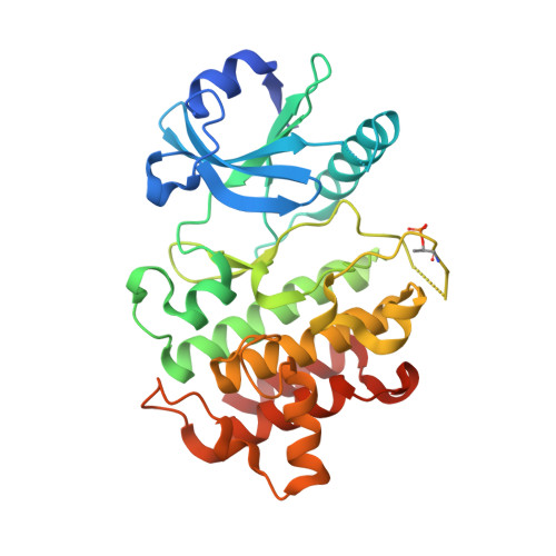
    Interleukin-1 (IL-1) receptor-associated kinase-4 (IRAK-4) is a serine/threonine kinase that plays an essential role in signal transduction by Toll/IL-1 receptors (TIRs). Here, we report the crystal structures of the phosphorylated human IRAK-4 kinase domain in complex with a potent inhibitor and with staurosporine to 2.0 and 2.2 A, respectively. The structures reveal that IRAK-4 has a unique tyrosine gatekeeper residue that interacts with the conserved glutamate from helix alphaC. Consequently, helix alphaC is "pulled in" to maintain the active orientation, and the usual pre-existing hydrophobic back pocket of the ATP-binding site is abolished. The peptide substrate-binding site is more open when compared with other protein kinases due to a marked movement of helix alphaG. The pattern of phosphate ligand interactions in the activation loop bears a close resemblance to that of a tyrosine kinase. Our results provide insights into IRAK-4 function and the design of selective inhibitors.


  • Organizational Affiliation

    Department of Molecular Structure, Amgen, Inc., 1120 Veterans Boulevard, South San Francisco, California 94080, USA. zwang@amgen.com


Macromolecules
Find similar proteins by:  (by identity cutoff)  |  3D Structure
Entity ID: 1
MoleculeChains Sequence LengthOrganismDetailsImage
interleukin-1 receptor-associated kinase 4
A, B, C, D
307Homo sapiensMutation(s): 1 
EC: 2.7.11.1
UniProt & NIH Common Fund Data Resources
Find proteins for Q9NWZ3 (Homo sapiens)
Explore Q9NWZ3 
Go to UniProtKB:  Q9NWZ3
PHAROS:  Q9NWZ3
GTEx:  ENSG00000198001 
Entity Groups  
Sequence Clusters30% Identity50% Identity70% Identity90% Identity95% Identity100% Identity
UniProt GroupQ9NWZ3
Sequence Annotations
Expand
  • Reference Sequence
Experimental Data & Validation

Experimental Data

  • Method: X-RAY DIFFRACTION
  • Resolution: 2.15 Å
  • R-Value Free: 0.261 
  • R-Value Work: 0.209 
  • R-Value Observed: 0.212 
  • Space Group: C 1 2 1
Unit Cell:
Length ( Å )Angle ( ˚ )
a = 141.502α = 90
b = 139.748β = 123.76
c = 87.656γ = 90
Software Package:
Software NamePurpose
REFMACrefinement

Structure Validation

View Full Validation Report


Ligand Structure Quality Assessment 


Entry History 

Deposition Data

Revision History  (Full details and data files)

  • Version 1.0: 2006-12-12
    Type: Initial release
  • Version 1.1: 2008-05-01
    Changes: Version format compliance
  • Version 1.2: 2011-07-13
    Changes: Version format compliance
  • Version 1.3: 2023-12-27
    Changes: Data collection, Database references, Derived calculations