2NOS

MURINE INDUCIBLE NITRIC OXIDE SYNTHASE OXYGENASE DOMAIN (DELTA 114), AMINOGUANIDINE COMPLEX


Experimental Data Snapshot

  • Method: X-RAY DIFFRACTION
  • Resolution: 2.30 Å
  • R-Value Free: 0.267 
  • R-Value Work: 0.204 
  • R-Value Observed: 0.204 

wwPDB Validation   3D Report Full Report


This is version 1.3 of the entry. See complete history


Literature

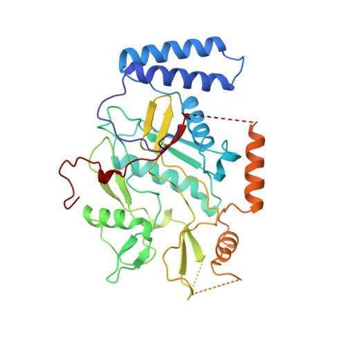
The structure of nitric oxide synthase oxygenase domain and inhibitor complexes.

Crane, B.R.Arvai, A.S.Gachhui, R.Wu, C.Ghosh, D.K.Getzoff, E.D.Stuehr, D.J.Tainer, J.A.

(1997) Science 278: 425-431

  • DOI: https://doi.org/10.1126/science.278.5337.425
  • Primary Citation of Related Structures:  
    1NOC, 1NOS, 2NOS

  • PubMed Abstract: 

    The nitric oxide synthase oxygenase domain (NOSox) oxidizes arginine to synthesize the cellular signal and defensive cytotoxin nitric oxide (NO). Crystal structures determined for cytokine-inducible NOSox reveal an unusual fold and heme environment for stabilization of activated oxygen intermediates key for catalysis. A winged beta sheet engenders a curved alpha-beta domain resembling a baseball catcher's mitt with heme clasped in the palm. The location of exposed hydrophobic residues and the results of mutational analysis place the dimer interface adjacent to the heme-binding pocket. Juxtaposed hydrophobic O2- and polar L-arginine-binding sites occupied by imidazole and aminoguanidine, respectively, provide a template for designing dual-function inhibitors and imply substrate-assisted catalysis.


  • Organizational Affiliation

    Department of Molecular Biology and the Skaggs Institute for Chemical Biology, The Scripps Research Institute, La Jolla, CA 92037, USA.


Macromolecules
Find similar proteins by:  (by identity cutoff)  |  3D Structure
Entity ID: 1
MoleculeChains Sequence LengthOrganismDetailsImage
INDUCIBLE NITRIC OXIDE SYNTHASE388Mus musculusMutation(s): 0 
EC: 1.14.13.39
UniProt & NIH Common Fund Data Resources
Find proteins for P29477 (Mus musculus)
Explore P29477 
Go to UniProtKB:  P29477
IMPC:  MGI:97361
Entity Groups  
Sequence Clusters30% Identity50% Identity70% Identity90% Identity95% Identity100% Identity
UniProt GroupP29477
Sequence Annotations
Expand
  • Reference Sequence
Small Molecules
Ligands 3 Unique
IDChains Name / Formula / InChI Key2D Diagram3D Interactions
HEM
Query on HEM

Download Ideal Coordinates CCD File 
B [auth A]PROTOPORPHYRIN IX CONTAINING FE
C34 H32 Fe N4 O4
KABFMIBPWCXCRK-RGGAHWMASA-L
AGU
Query on AGU

Download Ideal Coordinates CCD File 
D [auth A]AMINOGUANIDINE
C H6 N4
HAMNKKUPIHEESI-UHFFFAOYSA-N
IMD
Query on IMD

Download Ideal Coordinates CCD File 
C [auth A]IMIDAZOLE
C3 H5 N2
RAXXELZNTBOGNW-UHFFFAOYSA-O
Binding Affinity Annotations 
IDSourceBinding Affinity
AGU BindingDB:  2NOS IC50: min: 3900, max: 1.32e+5 (nM) from 8 assay(s)
Experimental Data & Validation

Experimental Data

  • Method: X-RAY DIFFRACTION
  • Resolution: 2.30 Å
  • R-Value Free: 0.267 
  • R-Value Work: 0.204 
  • R-Value Observed: 0.204 
  • Space Group: P 21 21 21
Unit Cell:
Length ( Å )Angle ( ˚ )
a = 63α = 90
b = 73.8β = 90
c = 92.8γ = 90
Software Package:
Software NamePurpose
X-PLORmodel building
X-PLORrefinement
DENZOdata reduction
SCALEPACKdata scaling
X-PLORphasing

Structure Validation

View Full Validation Report


Entry History 

Deposition Data

Revision History  (Full details and data files)

  • Version 1.0: 1998-10-14
    Type: Initial release
  • Version 1.1: 2008-03-24
    Changes: Version format compliance
  • Version 1.2: 2011-07-13
    Changes: Version format compliance
  • Version 1.3: 2024-02-21
    Changes: Data collection, Database references, Derived calculations, Other