2I9B

Crystal structure of ATF-urokinase receptor complex


Experimental Data Snapshot

  • Method: X-RAY DIFFRACTION
  • Resolution: 2.80 Å
  • R-Value Free: 0.265 
  • R-Value Work: 0.222 
  • R-Value Observed: 0.224 

wwPDB Validation   3D Report Full Report


This is version 2.2 of the entry. See complete history


Literature

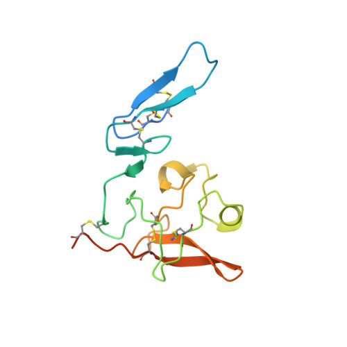
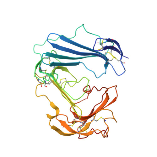
Structural basis of interaction between urokinase-type plasminogen activator and its receptor.

Barinka, C.Parry, G.Callahan, J.Shaw, D.E.Kuo, A.Bdeir, K.Cines, D.B.Mazar, A.Lubkowski, J.

(2006) J Mol Biol 363: 482-495

  • DOI: https://doi.org/10.1016/j.jmb.2006.08.063
  • Primary Citation of Related Structures:  
    2I9A, 2I9B

  • PubMed Abstract: 

    Recent studies indicate that binding of the urokinase-type plasminogen activator (uPA) to its high-affinity receptor (uPAR) orchestrates uPAR interactions with other cellular components that play a pivotal role in diverse (patho-)physiological processes, including wound healing, angiogenesis, inflammation, and cancer metastasis. However, notwithstanding the wealth of biochemical data available describing the activities of uPAR, little is known about the exact mode of uPAR/uPA interactions or the presumed conformational changes that accompany uPA/uPAR engagement. Here, we report the crystal structure of soluble urokinase plasminogen activator receptor (suPAR), which contains the three domains of the wild-type receptor but lacks the cell-surface anchoring sequence, in complex with the amino-terminal fragment of urokinase-type plasminogen activator (ATF), at the resolution of 2.8 A. We report the 1.9 A crystal structure of free ATF. Our results provide a structural basis, represented by conformational changes induced in uPAR, for several published biochemical observations describing the nature of uPAR/uPA interactions and provide insight into mechanisms that may be responsible for the cellular responses induced by uPA binding.


  • Organizational Affiliation

    Center for Cancer Research, National Cancer Institute, Frederick, MD 21702, USA.


Macromolecules
Find similar proteins by:  (by identity cutoff)  |  3D Structure
Entity ID: 1
MoleculeChains Sequence LengthOrganismDetailsImage
Urokinase-type plasminogen activator
A, B, C, D
145Homo sapiensMutation(s): 0 
Gene Names: PLAU
EC: 3.4.21.73
UniProt & NIH Common Fund Data Resources
Find proteins for P00749 (Homo sapiens)
Explore P00749 
Go to UniProtKB:  P00749
PHAROS:  P00749
GTEx:  ENSG00000122861 
Entity Groups  
Sequence Clusters30% Identity50% Identity70% Identity90% Identity95% Identity100% Identity
UniProt GroupP00749
Sequence Annotations
Expand
  • Reference Sequence
Find similar proteins by:  (by identity cutoff)  |  3D Structure
Entity ID: 2
MoleculeChains Sequence LengthOrganismDetailsImage
Urokinase plasminogen activator surface receptor
E, F, G, H
279Homo sapiensMutation(s): 4 
Gene Names: PLAURMO3UPAR
UniProt & NIH Common Fund Data Resources
Find proteins for Q03405 (Homo sapiens)
Explore Q03405 
Go to UniProtKB:  Q03405
PHAROS:  Q03405
GTEx:  ENSG00000011422 
Entity Groups  
Sequence Clusters30% Identity50% Identity70% Identity90% Identity95% Identity100% Identity
UniProt GroupQ03405
Sequence Annotations
Expand
  • Reference Sequence
Oligosaccharides

Help

Entity ID: 3
MoleculeChains Length2D Diagram Glycosylation3D Interactions
beta-D-mannopyranose-(1-6)-2-acetamido-2-deoxy-beta-D-glucopyranose-(1-4)-2-acetamido-2-deoxy-beta-D-glucopyranose
I, J
3N-Glycosylation
Glycosylation Resources
GlyTouCan:  G73474XW
GlyCosmos:  G73474XW
GlyGen:  G73474XW
Entity ID: 4
MoleculeChains Length2D Diagram Glycosylation3D Interactions
2-acetamido-2-deoxy-beta-D-glucopyranose-(1-4)-2-acetamido-2-deoxy-beta-D-glucopyranose
K, L
2N-Glycosylation
Glycosylation Resources
GlyTouCan:  G42666HT
GlyCosmos:  G42666HT
GlyGen:  G42666HT
Experimental Data & Validation

Experimental Data

  • Method: X-RAY DIFFRACTION
  • Resolution: 2.80 Å
  • R-Value Free: 0.265 
  • R-Value Work: 0.222 
  • R-Value Observed: 0.224 
  • Space Group: P 1 21 1
Unit Cell:
Length ( Å )Angle ( ˚ )
a = 62.914α = 90
b = 281.921β = 105.41
c = 62.811γ = 90
Software Package:
Software NamePurpose
REFMACrefinement
MAR345data collection
HKL-2000data reduction
HKL-2000data scaling
PHASERphasing

Structure Validation

View Full Validation Report


Entry History 

Deposition Data

Revision History  (Full details and data files)

  • Version 1.0: 2007-01-02
    Type: Initial release
  • Version 1.1: 2008-05-01
    Changes: Version format compliance
  • Version 1.2: 2011-07-13
    Changes: Advisory, Version format compliance
  • Version 1.3: 2017-10-18
    Changes: Advisory, Refinement description
  • Version 2.0: 2020-07-29
    Type: Remediation
    Reason: Carbohydrate remediation
    Changes: Advisory, Atomic model, Data collection, Database references, Derived calculations, Structure summary
  • Version 2.1: 2021-10-20
    Changes: Database references, Structure summary
  • Version 2.2: 2023-08-30
    Changes: Data collection, Refinement description