2HCI

Structure of Human Mip-3a Chemokine


Experimental Data Snapshot

  • Method: X-RAY DIFFRACTION
  • Resolution: 1.81 Å
  • R-Value Free: 0.255 
  • R-Value Work: 0.206 
  • R-Value Observed: 0.209 

wwPDB Validation   3D Report Full Report


This is version 1.3 of the entry. See complete history


Literature

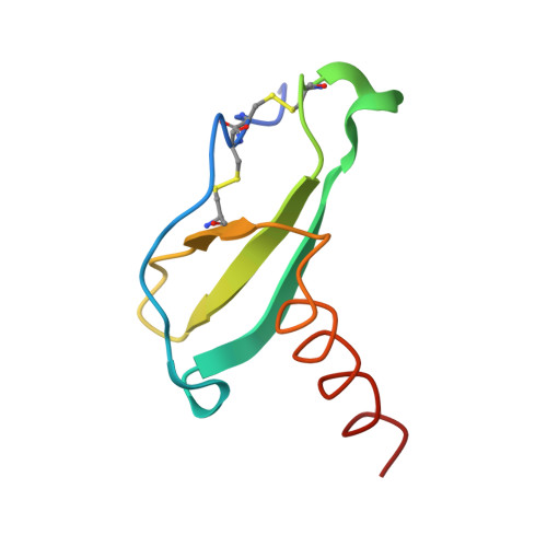
Structure of human MIP-3alpha chemokine.

Malik, Z.A.Tack, B.F.

(2006) Acta Crystallogr Sect F Struct Biol Cryst Commun 62: 631-634

  • DOI: https://doi.org/10.1107/S1744309106006890
  • Primary Citation of Related Structures:  
    2HCI

  • PubMed Abstract: 

    The structure of the human macrophage inflammatory protein-3alpha (MIP-3alpha) has been determined at 1.81 angstroms resolution by X-ray crystallography. The dimer crystallized in the tetragonal space group I4, with unit-cell parameters a = b = 83.99, c = 57.20 angstroms. The crystals exhibit two molecules in the asymmetric unit. The structure was solved by the molecular-replacement method and the model was refined to a conventional R value of 20.6% (R(free) = 25.7%). MIP-3alpha possesses the same monomeric structure as previously described for other chemokines. However, in addition to limited structural changes in the beta1-beta2 hairpin of monomer B, the electron density is fully defined for a few extra residues at the N- and C-termini of monomer A and the C-terminus of monomer B compared with MIP-3alpha in space group P6(1). As the N-terminal and loop regions have been shown to be critical for receptor binding and signaling, this additional structural information may help in determining the basis of the CCR6 selectivity of MIP-3alpha.


  • Organizational Affiliation

    Department of Pharmacology, Carver College of Medicine, The University of Iowa, Iowa City, IA 52242, USA. z-malik@uiowa.edu


Macromolecules
Find similar proteins by:  (by identity cutoff)  |  3D Structure
Entity ID: 1
MoleculeChains Sequence LengthOrganismDetailsImage
Small inducible cytokine A20
A, B
69N/AMutation(s): 0 
UniProt & NIH Common Fund Data Resources
Find proteins for P78556 (Homo sapiens)
Explore P78556 
Go to UniProtKB:  P78556
PHAROS:  P78556
GTEx:  ENSG00000115009 
Entity Groups  
Sequence Clusters30% Identity50% Identity70% Identity90% Identity95% Identity100% Identity
UniProt GroupP78556
Sequence Annotations
Expand
  • Reference Sequence
Small Molecules
Ligands 3 Unique
IDChains Name / Formula / InChI Key2D Diagram3D Interactions
PEG
Query on PEG

Download Ideal Coordinates CCD File 
H [auth A],
I [auth A],
J [auth B],
K [auth B]
DI(HYDROXYETHYL)ETHER
C4 H10 O3
MTHSVFCYNBDYFN-UHFFFAOYSA-N
SO4
Query on SO4

Download Ideal Coordinates CCD File 
C [auth A]SULFATE ION
O4 S
QAOWNCQODCNURD-UHFFFAOYSA-L
SO3
Query on SO3

Download Ideal Coordinates CCD File 
D [auth A],
E [auth A],
F [auth A],
G [auth A]
SULFITE ION
O3 S
LSNNMFCWUKXFEE-UHFFFAOYSA-L
Experimental Data & Validation

Experimental Data

  • Method: X-RAY DIFFRACTION
  • Resolution: 1.81 Å
  • R-Value Free: 0.255 
  • R-Value Work: 0.206 
  • R-Value Observed: 0.209 
  • Space Group: I 4
Unit Cell:
Length ( Å )Angle ( ˚ )
a = 83.991α = 90
b = 83.991β = 90
c = 57.221γ = 90
Software Package:
Software NamePurpose
DENZOdata reduction
d*TREKdata reduction
AMoREphasing
REFMACrefinement

Structure Validation

View Full Validation Report


Entry History 

Deposition Data

Revision History  (Full details and data files)

  • Version 1.0: 2006-07-04
    Type: Initial release
  • Version 1.1: 2008-05-01
    Changes: Version format compliance
  • Version 1.2: 2011-07-13
    Changes: Version format compliance
  • Version 1.3: 2023-08-30
    Changes: Data collection, Database references, Derived calculations, Refinement description