The RNA recognition mechanism of human immunodeficiency virus (HIV) type 2 NCp8 is different from that of HIV-1 NCp7
Matsui, T., Tanaka, T., Endoh, H., Sato, K., Tanaka, H., Miyauchi, E., Kawashima, Y., Nagai-Makabe, M., Komatsu, H., Kohno, T., Maeda, T., Kodera, Y.(2009) Biochemistry 48: 4314-4323
- PubMed: 19334676
- DOI: https://doi.org/10.1021/bi802364b
- Primary Citation of Related Structures:
2EC7 - PubMed Abstract:
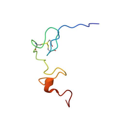
The nucleocapsid (NC) protein of HIV, which contains two CCHC-type zinc fingers connected by a linker, is a multifunctional protein involved in many of the critical steps of the HIV life cycle. HIV-1 and HIV-2 contain NC proteins NCp7 and NCp8, respectively. The amino acid sequences of both NC proteins are 67% identical. For NCp7, the important elements for RNA binding were found to be the first zinc finger flanked by the linker, as the minimal active domain, and the 3(10) helix in the N-terminus, as the secondary active domain. However, for the NCp8 counterpart in HIV-2, the mechanism for binding to viral RNA has not yet been clarified. In this study, we determined NCp8's three-dimensional structure for the first time and examined the dynamic behavior and chemical shift perturbation as a function of the concentration of viral RNA SL3. Moreover, the specific binding activities of NCp8 and the NCp8-derived peptides with SL3 were examined by a native polyacrylamide gel electrophoresis assay. These results indicate that the RNA recognition mechanism for NCp8 is different from that of NCp7 and that the hydrophobic cleft in the second zinc finger acts as a secondary active domain instead of the 3(10) helix in NCp7. Furthermore, the flexibility of the linker is limited by the hydrogen bond between the first zinc finger (Asn11) and the linker (Arg27), which makes it possible for the sites around Trp10 in the minimal active domain and the secondary active domain to form the binding surface.
Organizational Affiliation:
Department of Physics, School of Science, Kitasato University, Sagamihara, Kanagawa 228-8555, Japan.