2DU2

Crystal Structure Analysis of the L-Lactate Oxidase


Experimental Data Snapshot

  • Method: X-RAY DIFFRACTION
  • Resolution: 2.10 Å
  • R-Value Free: 0.199 
  • R-Value Work: 0.160 
  • R-Value Observed: 0.162 

wwPDB Validation   3D Report Full Report


Ligand Structure Quality Assessment 


This is version 1.3 of the entry. See complete history


Literature

The crystal structure of L-lactate oxidase from Aerococcus viridans at 2.1A resolution reveals the mechanism of strict substrate recognition

Umena, Y.Yorita, K.Matsuoka, T.Kita, A.Fukui, K.Morimoto, Y.

(2006) Biochem Biophys Res Commun 350: 249-256

  • DOI: https://doi.org/10.1016/j.bbrc.2006.09.025
  • Primary Citation of Related Structures:  
    2DU2

  • PubMed Abstract: 

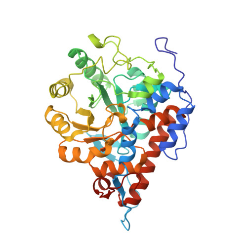
    L-Lactate oxidase (LOX) from Aerococcus viridans is a member of the alpha-hydroxyacid-oxidase flavoenzyme family. We have determined the three-dimensional structure of LOX and revealed the mechanism of substrate recognition. The LOX monomer structure has a typical alpha(8)/beta(8) motif commonly found in other flavin family proteins. A related enzyme, glycolate oxidase, catalyzes the oxidation of glycolate rather than lactate. Comparison of the two enzyme structures highlights the importance of five residues around the FMN prosthetic group of LOX, which act synergistically to discriminate between the l/d configurations of lactate. X-ray crystallography of LOX gave a space group I422 of unit-cell parameters a=b=191.096A, c=194.497A and alpha=beta=gamma=90 degrees with four monomers per asymmetric unit. The four independent monomers display slight structural differences around the active site. Diffraction data were collected, under cryogenic conditions to 2.1A resolution at the synchrotron facilities in Japan.


  • Organizational Affiliation

    Research Reactor Institute, Kyoto University, Kumatori, Osaka 590-0494, Japan.


Macromolecules
Find similar proteins by:  (by identity cutoff)  |  3D Structure
Entity ID: 1
MoleculeChains Sequence LengthOrganismDetailsImage
Lactate oxidase
A, B, C, D
374Aerococcus viridansMutation(s): 0 
EC: 1.13.12.4
UniProt
Find proteins for Q44467 (Aerococcus viridans (strain ATCC 11563 / DSM 20340 / CCUG 4311 / JCM 20461 / NBRC 12219 / NCTC 8251 / M1))
Explore Q44467 
Go to UniProtKB:  Q44467
Entity Groups  
Sequence Clusters30% Identity50% Identity70% Identity90% Identity95% Identity100% Identity
UniProt GroupQ44467
Sequence Annotations
Expand
  • Reference Sequence
Experimental Data & Validation

Experimental Data

  • Method: X-RAY DIFFRACTION
  • Resolution: 2.10 Å
  • R-Value Free: 0.199 
  • R-Value Work: 0.160 
  • R-Value Observed: 0.162 
  • Space Group: I 4 2 2
Unit Cell:
Length ( Å )Angle ( ˚ )
a = 191.096α = 90
b = 191.096β = 90
c = 194.497γ = 90
Software Package:
Software NamePurpose
REFMACrefinement
HKL-2000data reduction
SCALEPACKdata scaling
AMoREphasing

Structure Validation

View Full Validation Report


Ligand Structure Quality Assessment 


Entry History 

Deposition Data

  • Released Date: 2006-12-05 
  • Deposition Author(s): Morimoto, Y.

Revision History  (Full details and data files)

  • Version 1.0: 2006-12-05
    Type: Initial release
  • Version 1.1: 2008-04-30
    Changes: Version format compliance
  • Version 1.2: 2011-07-13
    Changes: Version format compliance
  • Version 1.3: 2023-10-25
    Changes: Data collection, Database references, Derived calculations, Refinement description