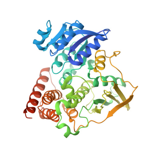
The 1.9 A Resolution Structure of Mycobacterium Tuberculosis 1-Deoxy-D-Xylulose 5-Phosphate Reductoisomerase, a Potential Drug Target.
Henriksson, L.M., Bjorkelid, C., Mowbray, S.L., Unge, T.(2006) Acta Crystallogr D Biol Crystallogr 62: 807
- PubMed: 16790937
- DOI: https://doi.org/10.1107/S0907444906019196
- Primary Citation of Related Structures:
2C82 - PubMed Abstract:
1-deoxy-D-xylulose 5-phosphate reductoisomerase catalyzes the NADPH-dependent rearrangement and reduction of 1-deoxy-D-xylulose 5-phosphate to form 2-C-methyl-D-erythritol 4-phosphate, as the second step of the deoxyxylulose 5-phosphate/methylerythritol 4-phosphate pathway found in many bacteria and plants. The end product, isopentenyl diphosphate, is the precursor of various isoprenoids vital to all living organisms. The pathway is not found in humans; the mevalonate pathway is instead used for the formation of isopentenyl diphosphate. This difference, combined with its essentiality, makes the reductoisomerase an excellent drug target in a number of pathogenic organisms. The structure of 1-deoxy-D-xylulose 5-phosphate reductoisomerase from Mycobacterium tuberculosis (Rv2870c) was solved by molecular replacement and refined to a resolution of 1.9 A. The enzyme exhibited an estimated kcat of 5.3 s-1 and Km and kcat/Km values of 7.2 microM and 7.4x10(5) M-1 s-1 for NADPH and 340 microM and 1.6x10(4) M-1 s-1 for 1-deoxy-D-xylulose 5-phosphate. In the structure, a sulfate is bound at the expected site of the phosphate moiety of the sugar substrate. The M. tuberculosis enzyme displays a similar fold to the previously published structures from Escherichia coli and Zymomonas mobilis. Comparisons offer suggestions for the design of specific drugs. Furthermore, the new structure represents an intermediate conformation between the open apo form and the closed holo form observed previously, giving insights into the conformational changes associated with catalysis.
Organizational Affiliation:
Department of Cell and Molecular Biology, Uppsala University, Biomedical Center, Box 596, SE-751 24 Uppsala, Sweden.