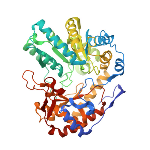
Structure of Escherichia Coli Tryptophanase
Ku, S.-Y., Yip, P., Howell, P.L.(2006) Acta Crystallogr D Biol Crystallogr 62: 814
- PubMed: 16790938
- DOI: https://doi.org/10.1107/S0907444906019895
- Primary Citation of Related Structures:
2C44 - PubMed Abstract:
Pyridoxal 5'-phosphate (PLP) dependent tryptophanase has been isolated from Escherichia coli and its crystal structure has been determined. The structure shares the same fold with and has similar quaternary structure to Proteus vulgaris tryptophanase and tyrosine-phenol lyase, but is found in a closed conformation when compared with these two enzymes. The tryptophanase structure, solved in its apo form, does not have covalent PLP bound in the active site, but two sulfate ions. The sulfate ions occupy the phosphoryl-binding site of PLP and the binding site of the alpha-carboxyl of the natural substrate tryptophan. One of the sulfate ions makes extensive interactions with both the transferase and PLP-binding domains of the protein and appears to be responsible for holding the enzyme in its closed conformation. Based on the sulfate density and the structure of the P. vulgaris enzyme, PLP and the substrate tryptophan were modeled into the active site. The resulting model is consistent with the roles of Arg419 in orienting the substrate to PLP and acidifying the alpha-proton of the substrate for beta-elimination, Lys269 in the formation and decomposition of the PLP quinonoid intermediate, Arg230 in orienting the substrate-PLP intermediates in the optimal conformation for catalysis, and His463 and Tyr74 in determining substrate specificity and suggests that the closed conformation observed in the structure could be induced by substrate binding and that significant conformational changes occur during catalysis. A catalytic mechanism for tryptophanase is proposed. Since E. coli tryptophanase has resisted forming diffraction-quality crystals for many years, the molecular surface of tryptophanase has been analyzed in various crystal forms and it was rationalized that strong crystal contacts occur on the flat surface of the protein and that the size of crystal contact surface seems to correlate with the diffraction quality of the crystal.
Organizational Affiliation:
Structural Biology and Biochemistry, Research Institute, Hospital for Sick Children, and Department of Biochemistry, Faculty of Medicine, University of Toronto, Ontario M5G 1X8, Canada.