Inter-domain motions of the N-domain of the KdpFABC complex, a P-type ATPase, are not driven by ATP-induced conformational changes.
Haupt, M., Bramkamp, M., Coles, M., Altendorf, K., Kessler, H.(2004) J Mol Biol 342: 1547-1558
- PubMed: 15364580
- DOI: https://doi.org/10.1016/j.jmb.2004.07.060
- Primary Citation of Related Structures:
1SVJ, 1U7Q - PubMed Abstract:
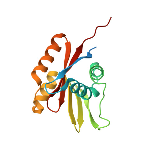
P-type ATPases are involved in the active transport of ions across biological membranes. The KdpFABC complex (P-type ATPase) of Escherichia coli is a high-affinity K+ uptake system that operates only when the cell experiences osmotic stress or K+ limitation. Here, we present the solution structure of the nucleotide binding domain of KdpB (backbone RMSD 0.17 A) and a model of the AMP-PNP binding mode based on intermolecular distance restraints. The calculated AMP-PNP binding mode shows the purine ring of the nucleotide to be "clipped" into the binding pocket via a pi-pi-interaction to F377 on one side and a cation-pi-interaction to K395 on the other. This binding mechanism seems to be conserved in all P-type ATPases, except the heavy metal transporting ATPases (type IB). Thus, we conclude that the Kdp-ATPase (currently type IA) is misgrouped and has more similarities to type III ATPases. The KdpB N-domain is the smallest and simplest known for a P-type ATPase, and represents a minimal example of this functional unit. No evidence of significant conformational changes was observed within the N-domain upon nucleotide binding, thus ruling out a role for ATP-induced conformational changes in the reaction cycle.
Organizational Affiliation:
Institut für Organische Chemie und Biochemie, Technische Universität München, Lichtenbergstr. 4, 85747 Garching, Germany.