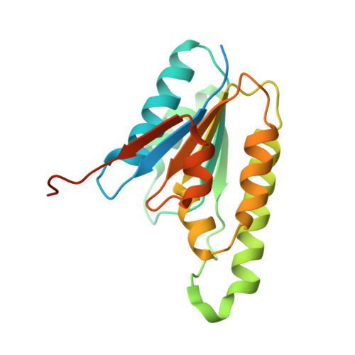
Three-dimensional structure of ATP:corrinoid adenosyltransferase from Salmonella typhimurium in its free state, complexed with MgATP, or complexed with hydroxycobalamin and MgATP.
Bauer, C.B., Fonseca, M.V., Holden, H.M., Thoden, J.B., Thompson, T.B., Escalante-Semerena, J.C., Rayment, I.(2001) Biochemistry 40: 361-374
- PubMed: 11148030
- DOI: https://doi.org/10.1021/bi002145o
- Primary Citation of Related Structures:
1G5R, 1G5T, 1G64 - PubMed Abstract:
In Salmonella typhimurium, formation of the cobalt-carbon bond in the biosynthetic pathway for adenosylcobalamin is catalyzed by the product of the cobA gene which encodes a protein of 196 amino acid residues. This enzyme is an ATP:co(I)rrinoid adenosyltransferase which transfers an adenosyl moiety from MgATP to a broad range of co(I)rrinoid substrates that are believed to include cobinamide, its precursor cobyric acid and probably others as yet unidentified, and hydroxocobalamin. Three X-ray structures of CobA are reported here: its substrate-free form, a complex of CobA with MgATP, and a ternary complex of CobA with MgATP and hydroxycobalamin to 2.1, 1.8, and 2.1 A resolution, respectively. These structures show that the enzyme is a homodimer. In the apo structure, the polypeptide chain extends from Arg(28) to Lys(181) and consists of an alpha/beta structure built from a six-stranded parallel beta-sheet with strand order 324516. The topology of this fold is very similar to that seen in RecA protein, helicase domain, F(1)ATPase, and adenosylcobinamide kinase/adenosylcobinamide guanylyltransferase where a P-loop is located at the end of the first strand. Strikingly, the nucleotide in the MgATP.CobA complex binds to the P-loop of CobA in the opposite orientation compared to all the other nucleotide hydrolases. That is, the gamma-phosphate binds at the location normally occupied by the alpha-phosphate. The unusual orientation of the nucleotide arises because this enzyme transfers an adenosyl group rather than the gamma-phosphate. In the ternary complex, the binding site for hydroxycobalamin is located in a shallow bowl-shaped depression at the C-terminal end of the beta-sheet of one subunit; however, the active site is capped by the N-terminal helix from the symmetry-related subunit that now extends from Gln(7) to Ala(24). The lower ligand of cobalamin is well-ordered and interacts mostly with the N-terminal helix of the symmetry-related subunit. Interestingly, there are few interactions between the protein and the polar side chains of the corrin ring which accounts for the broad specificity of this enzyme. The corrin ring is oriented such that the cobalt atom is located approximately 6.1 A from C5' of the ribose and is beyond the range of nucleophilic attack. This suggests that a conformational change occurs in the ternary complex when Co(III) is reduced to Co(I).
Organizational Affiliation:
Department of Biochemistry, University of Wisconsin, Madison, Wisconsin 53706, USA.