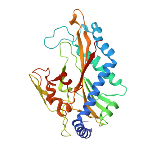
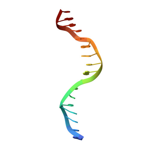
1DMU

Crystal structure of the restriction endonuclease BglI (e.c.3.1.21.4) bound to its dna recognition sequence


Experimental Data Snapshot

  • Method: X-RAY DIFFRACTION
  • Resolution: 2.20 Å
  • R-Value Free: 0.239 
  • R-Value Work: 0.177 
  • R-Value Observed: 0.183 

wwPDB Validation   3D Report Full Report


This is version 1.3 of the entry. See complete history


Literature

Crystal structure of restriction endonuclease BglI bound to its interrupted DNA recognition sequence.

Newman, M.Lunnen, K.Wilson, G.Greci, J.Schildkraut, I.Phillips, S.E.

(1998) EMBO J 17: 5466-5476

  • DOI: https://doi.org/10.1093/emboj/17.18.5466
  • Primary Citation of Related Structures:  
    1DMU

  • PubMed Abstract: 

    The crystal structure of the type II restriction endonuclease BglI bound to DNA containing its specific recognition sequence has been determined at 2.2 A resolution. This is the first structure of a restriction endonuclease that recognizes and cleaves an interrupted DNA sequence, producing 3' overhanging ends. BglI is a homodimer that binds its specific DNA sequence with the minor groove facing the protein. Parts of the enzyme reach into both the major and minor grooves to contact the edges of the bases within the recognition half-sites. The arrangement of active site residues is strikingly similar to other restriction endonucleases, but the co-ordination of two calcium ions at the active site gives new insight into the catalytic mechanism. Surprisingly, the core of a BglI subunit displays a striking similarity to subunits of EcoRV and PvuII, but the dimer structure is dramatically different. The BglI-DNA complex demonstrates, for the first time, that a conserved subunit fold can dimerize in more than one way, resulting in different DNA cleavage patterns.


  • Organizational Affiliation

    School of Biochemistry and Molecular Biology, and North of England Structural Biology Centre, University of Leeds, Leeds LS2 9JT, UK.


Macromolecules

Find similar proteins by:  (by identity cutoff)  |  3D Structure
Entity ID: 2
MoleculeChains Sequence LengthOrganismDetailsImage
BGLI RESTRICTION ENDONUCLEASEB [auth A]299Bacillus subtilisMutation(s): 0 
EC: 3.1.21.4
UniProt
Find proteins for O68557 (Bacillus subtilis)
Explore O68557 
Go to UniProtKB:  O68557
Entity Groups  
Sequence Clusters30% Identity50% Identity70% Identity90% Identity95% Identity100% Identity
UniProt GroupO68557
Sequence Annotations
Expand
  • Reference Sequence

Find similar nucleic acids by:  Sequence   |   3D Structure  

Entity ID: 1
MoleculeChains LengthOrganismImage
DNA (5'-D(*AP*TP*CP*GP*CP*CP*TP*AP*AP*TP*AP*GP*GP*CP*GP*AP*T)-3')A [auth F]17N/A
Sequence Annotations
Expand
  • Reference Sequence
Experimental Data & Validation

Experimental Data

  • Method: X-RAY DIFFRACTION
  • Resolution: 2.20 Å
  • R-Value Free: 0.239 
  • R-Value Work: 0.177 
  • R-Value Observed: 0.183 
  • Space Group: C 2 2 21
Unit Cell:
Length ( Å )Angle ( ˚ )
a = 78.48α = 90
b = 81.6β = 90
c = 117.06γ = 90
Software Package:
Software NamePurpose
SHARPphasing
X-PLORrefinement
DENZOdata reduction
SCALEPACKdata scaling

Structure Validation

View Full Validation Report


Entry History 

Deposition Data

Revision History  (Full details and data files)

  • Version 1.0: 1999-12-18
    Type: Initial release
  • Version 1.1: 2008-04-27
    Changes: Version format compliance
  • Version 1.2: 2011-07-13
    Changes: Version format compliance
  • Version 1.3: 2021-02-03
    Changes: Database references, Derived calculations