2'-Deoxyisoguanosine adopts more than one tautomer to form base pairs with thymidine observed by high-resolution crystal structure analysis.
Robinson, H., Gao, Y.G., Bauer, C., Roberts, C., Switzer, C., Wang, A.H.(1998) Biochemistry 37: 10897-10905
- PubMed: 9692982
- DOI: https://doi.org/10.1021/bi980818l
- Primary Citation of Related Structures:
1BHR, 403D - PubMed Abstract:
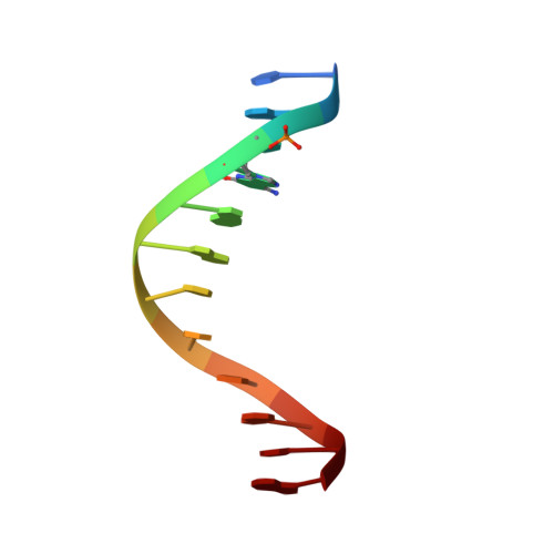
The questions of whether different tautomeric forms of nucleic acid bases exist to any significant extent in DNA, or what their possible roles in mutation may be, are under intense scrutiny. 2'-Deoxyisoguanosine (iG) has been suggested to have a propensity to adopt the enol form. Isoguanine (also called 2-hydroxyadenine) can be found in oxidatively damaged DNA generated from treating DNA with a Fenton-type reactive oxygen-generating system and is known to cause mutation. We have analyzed the three-dimensional structure of the DNA dodecamer d(CGC[iG]AATTTGCG) (denoted iG-DODE) by X-ray crystallography and NMR. The crystal structure of the iG-DODE complexed with the minor groove binder Hoechst 33342, refined to 1.4 A resolution, showed that the two independent iG.T base pairs in the dodecamer duplex adopt different (one in Watson-Crick and the other in wobble) conformations. The high-resolution nature of the structure also affords unprecedented clear information about the conformation and interactions of the Hoechst drug. The Hoechst 33342 binds in the narrow minor groove at the iGAATT site, with the N-methylpiperazine ring near the iG4.T21 base pair. Three hydrogen bonds are found between the NH of the Hoechst ligand and T-O2 DNA atoms. In solution, the two iG.T base pairs in iG-DODE predominantly are in the wobble form at 2 degreesC. At higher temperatures, another duplex form (likely involving the enol form of iG) is in slow exchange with the keto form and becomes significantly populated, reaching approximately 40% at 40 degreesC. Our data support the conclusion that iG pairs with T in a Watson-Crick configuration to a significant extent at physiological temperature (37 degreesC), which may explain the facile incorporation rate of T across from an iG during in vitro DNA replication.
Organizational Affiliation:
Department of Cell and Structural Biology, University of Illinois at Urbana-Champaign 61801, USA.