1ZCD

Crystal structure of the Na+/H+ antiporter NhaA


Experimental Data Snapshot

  • Method: X-RAY DIFFRACTION
  • Resolution: 3.45 Å
  • R-Value Free: 0.316 
  • R-Value Work: 0.316 
  • R-Value Observed: 0.302 

wwPDB Validation   3D Report Full Report


This is version 1.3 of the entry. See complete history


Literature

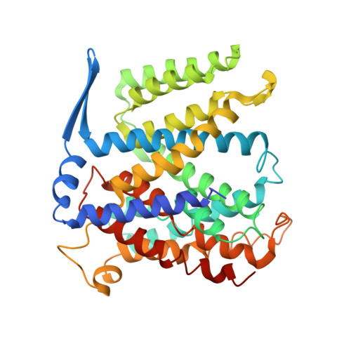
Structure of a Na+/H+ antiporter and insights into mechanism of action and regulation by pH.

Hunte, C.Screpanti, E.Venturi, M.Rimon, A.Padan, E.Michel, H.

(2005) Nature 435: 1197-1202

  • DOI: https://doi.org/10.1038/nature03692
  • Primary Citation of Related Structures:  
    1ZCD

  • PubMed Abstract: 

    The control by Na+/H+ antiporters of sodium/proton concentration and cell volume is crucial for the viability of all cells. Adaptation to high salinity and/or extreme pH in plants and bacteria or in human heart muscles requires the action of Na+/H+ antiporters. Their activity is tightly controlled by pH. Here we present the crystal structure of pH-downregulated NhaA, the main antiporter of Escherichia coli and many enterobacteria. A negatively charged ion funnel opens to the cytoplasm and ends in the middle of the membrane at the putative ion-binding site. There, a unique assembly of two pairs of short helices connected by crossed, extended chains creates a balanced electrostatic environment. We propose that the binding of charged substrates causes an electric imbalance, inducing movements, that permit a rapid alternating-access mechanism. This ion-exchange machinery is regulated by a conformational change elicited by a pH signal perceived at the entry to the cytoplasmic funnel.


  • Organizational Affiliation

    Department of Molecular Membrane Biology, Max Planck Institute of Biophysics, Max-von-Laue-Str. 3, D-60438 Frankfurt, Germany.


Macromolecules
Find similar proteins by:  (by identity cutoff)  |  3D Structure
Entity ID: 1
MoleculeChains Sequence LengthOrganismDetailsImage
Na(+)/H(+) antiporter 1
A, B
388Escherichia coliMutation(s): 0 
Gene Names: nhaA
Membrane Entity: Yes 
UniProt
Find proteins for P13738 (Escherichia coli (strain K12))
Explore P13738 
Go to UniProtKB:  P13738
Entity Groups  
Sequence Clusters30% Identity50% Identity70% Identity90% Identity95% Identity100% Identity
UniProt GroupP13738
Sequence Annotations
Expand
  • Reference Sequence
Experimental Data & Validation

Experimental Data

  • Method: X-RAY DIFFRACTION
  • Resolution: 3.45 Å
  • R-Value Free: 0.316 
  • R-Value Work: 0.316 
  • R-Value Observed: 0.302 
  • Space Group: P 21 21 21
Unit Cell:
Length ( Å )Angle ( ˚ )
a = 108.868α = 90
b = 121.718β = 90
c = 123.578γ = 90
Software Package:
Software NamePurpose
DENZOdata reduction
XDSdata reduction
SOLVEphasing
SHARPphasing
CNSrefinement
SCALEPACKdata scaling
XDSdata scaling

Structure Validation

View Full Validation Report


Entry History 

Deposition Data

Revision History  (Full details and data files)

  • Version 1.0: 2005-07-05
    Type: Initial release
  • Version 1.1: 2008-04-30
    Changes: Version format compliance
  • Version 1.2: 2011-07-13
    Changes: Version format compliance
  • Version 1.3: 2024-03-13
    Changes: Data collection, Database references