Structural and Functional Study of an Anemonia Elastase Inhibitor, a "Nonclassical" Kazal-Type Inhibitor from Anemonia sulcata
Hemmi, H., Kumazaki, T., Yoshizawa-Kumagaye, K., Nishiuchi, Y., Yoshida, T., Ohkubo, T., Kobayashi, Y.(2005) Biochemistry 44: 9626-9636
- PubMed: 16008348
- DOI: https://doi.org/10.1021/bi0472806
- Primary Citation of Related Structures:
1Y1B, 1Y1C - PubMed Abstract:
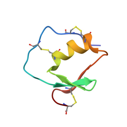
Anemonia elastase inhibitor (AEI) is a "nonclassical" Kazal-type elastase inhibitor from Anemonia sulcata. Unlike many nonclassical inhibitors, AEI does not have a cystine-stabilized alpha-helical (CSH) motif in the sequence. We chemically synthesized AEI and determined its three-dimensional solution structure by two-dimensional NMR spectroscopy. The resulting structure of AEI was characterized by a central alpha-helix and a three-stranded antiparallel beta-sheet of a typical Kazal-type inhibitor such as silver pheasant ovomucoid third domain (OMSVP3), even though the first and fifth half-cystine residues forming a disulfide bond in AEI are shifted both toward the C-terminus in comparison with those of OMSVP3. Synthesized AEI exhibited unexpected strong inhibition toward Streptomyces griseus protease B (SGPB). Our previous study [Hemmi, H., et al. (2003) Biochemistry 42, 2524-2534] demonstrated that the site-specific introduction of the engineered disulfide bond into the OMSVP3 molecule to form the CSH motif could produce an inhibitor with a narrower specificity. Thus, the CSH motif-containing derivative of AEI (AEI analogue) was chemically synthesized when a Cys(4)-Cys(34) bond was changed to a Cys(6)-Cys(31) bond. The AEI analogue scarcely inhibited porcine pancreatic elastase (PPE), even though it exhibited almost the same potent inhibitory activity toward SGPB. For the molecular scaffold, essentially no structural difference was detected between the two, but the N-terminal loop from Pro(5) to Ile(7) near the putative reactive site (Met(10)-Gln(11)) in the analogue moved by 3.7 A toward the central helix to form the introduced Cys(6)-Cys(31) bond. Such a conformational change in the restricted region correlates with the specificity change of the inhibitor.
Organizational Affiliation:
National Food Research Institute, 2-1-12 Kannondai, Tsukuba, Ibaraki 305-8642, Japan.