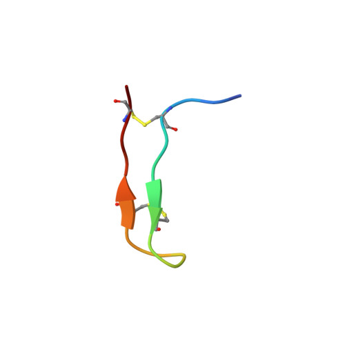
Solution structure and interaction of the antimicrobial polyphemusins with lipid membranes
Powers, J.P., Tan, A., Ramamoorthy, A., Hancock, R.E.(2005) Biochemistry 44: 15504-15513
- PubMed: 16300399
- DOI: https://doi.org/10.1021/bi051302m
- Primary Citation of Related Structures:
1X7K, 2B5K - PubMed Abstract:
The horseshoe crab cationic antimicrobial peptide polyphemusin I is highly active in vitro but not protective in mouse models of bacterial and LPS challenge, while a synthetic polyphemusin variant, PV5, was previously shown to be protective in vivo. In this study, we investigated the interaction of these peptides with lipid membranes in an effort to propose a mechanism of interaction. The solution structure of PV5 was determined by proton NMR in the absence and presence of dodecylphosphocholine (DPC) micelles. Like polyphemusin I, PV5 is a beta-hairpin but appeared less amphipathic in solution. Upon association with DPC micelles, PV5 underwent side chain rearrangements which resulted in an increased amphipathic conformation. Using fluorescence spectroscopy, both peptides were found to have limited affinity for neutral vesicles composed of phosphatidylcholine (PC). Incorporation of 25 mol % cholesterol or phosphatidylethanolamine into PC vesicles produced little change in the partitioning of either peptide. Incorporation of 25 mol % phosphatidylglycerol (PG) into PC vesicles, a simple prokaryotic model, resulted in a large increase in the affinity for both peptides, but the partition coefficient for PV5 was almost twice that of polyphemusin I. Differential scanning calorimetry studies supported the partitioning data and demonstrated that neither peptide interacted readily with neutral PC vesicles. Both peptides showed affinity for negatively charged membranes incorporating PG. The affinity of PV5 was much greater as the pretransition peak was absent at low peptide to lipid ratios (1:400) and the reduction in enthalpy of the main transition was greater than that produced by polyphemusin I. Both peptides decreased the lamellar to inverted hexagonal phase transition temperature of PE indicating the induction of negative curvature strain. These results, combined with previous findings that polyphemusin I promotes lipid flip-flop but does not induce significant vesicle leakage, ruled out the torroidal pore and carpet mechanisms of antimicrobial action for these polyphemusins.
Organizational Affiliation:
Department of Microbiology and Immunology, University of British Columbia, Vancouver, British Columbia V6T 1Z3, Canada.