


Crystal structure of Pyrococcus horikoshii phosphomannomutase/phosphoglucomutase
Kawamura, T., Tsuge, M., Watanabe, N., Tanaka, I.To be published.
Experimental Data Snapshot
wwPDB Validation 3D Report Full Report

Entity ID: 1 | |||||
|---|---|---|---|---|---|
| Molecule | Chains | Sequence Length | Organism | Details | Image |
| phospho-sugar mutase | 455 | Pyrococcus horikoshii | Mutation(s): 0 Gene Names: PH0923 EC: 5.4.2.8 | 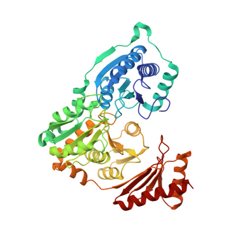 | |
UniProt | |||||
Find proteins for O58651 (Pyrococcus horikoshii (strain ATCC 700860 / DSM 12428 / JCM 9974 / NBRC 100139 / OT-3)) Explore O58651 Go to UniProtKB: O58651 | |||||
Entity Groups | |||||
| Sequence Clusters | 30% Identity50% Identity70% Identity90% Identity95% Identity100% Identity | ||||
| UniProt Group | O58651 | ||||
Sequence AnnotationsExpand | |||||
| |||||
| Ligands 1 Unique | |||||
|---|---|---|---|---|---|
| ID | Chains | Name / Formula / InChI Key | 2D Diagram | 3D Interactions | |
| MG Query on MG | E [auth A], F [auth B], G [auth C], H [auth D] | MAGNESIUM ION Mg JLVVSXFLKOJNIY-UHFFFAOYSA-N |  | ||
| Length ( Å ) | Angle ( ˚ ) |
|---|---|
| a = 77.653 | α = 90 |
| b = 137.916 | β = 93.27 |
| c = 98.409 | γ = 90 |
| Software Name | Purpose |
|---|---|
| DENZO | data reduction |
| SCALEPACK | data scaling |
| AMoRE | phasing |
| CNS | refinement |
RCSB PDB (citation) is hosted by
RCSB PDB is a member of the

