1WD0

Crystal structures of the hyperthermophilic chromosomal protein Sac7d in complex with DNA decamers


Experimental Data Snapshot

  • Method: X-RAY DIFFRACTION
  • Resolution: 1.90 Å
  • R-Value Free: 0.257 
  • R-Value Work: 0.221 
  • R-Value Observed: 0.224 

wwPDB Validation   3D Report Full Report


This is version 1.3 of the entry. See complete history


Literature

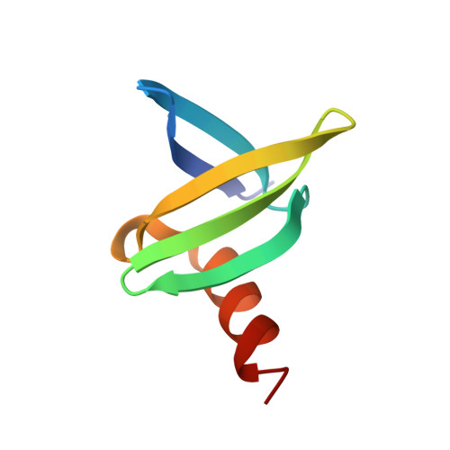
Structures of the hyperthermophilic chromosomal protein Sac7d in complex with DNA decamers.

Ko, T.P.Chu, H.M.Chen, C.Y.Chou, C.C.Wang, A.H.

(2004) Acta Crystallogr D Biol Crystallogr 60: 1381-1387

  • DOI: https://doi.org/10.1107/S0907444904012065
  • Primary Citation of Related Structures:  
    1WD0, 1WD1

  • PubMed Abstract: 

    The protein Sac7d belongs to a class of small chromosomal proteins from the hyperthermophilic archaeon Sulfolobus acidocaldarius. Two new crystal forms of Sac7d in complex with the DNA decamers CCTATATAGG and CCTACGTAGG were obtained and their structures were determined by molecular replacement. The refined models yielded R/Rfree values of 0.221/0.257 and 0.248/0.290 at 1.9 and 2.2 A resolution, respectively. The protein structures are similar to the previously determined structure of Sac7d-GCGATCGC (PDB code 1azp), but the DNA molecules are more bent overall, by 14-20 degrees. The relative positions of the Sac7d protein and the bound DNA also differ by rotations of 6-10 degrees and translations of 1.0-2.4 A. In addition to the water molecules in the central cavity, three additional conserved water molecules are found that mediate the protein-DNA interactions. The decamer DNA fragments form virtual double helices in the crystal, with a unit length of eight base pairs. The molecular packing of the new crystal forms differs from that of 1azp. The terminal nucleotides are opened up and form triple base pairs with other DNA molecules. Through lattice contacts, the Sac7d molecule also makes additional interactions with DNA, whereas only limited protein-protein interactions are seen.


  • Organizational Affiliation

    Institute of Biological Chemistry, Academia Sinica, Taipei 115, Taiwan.


Macromolecules

Find similar proteins by:  (by identity cutoff)  |  3D Structure
Entity ID: 2
MoleculeChains Sequence LengthOrganismDetailsImage
DNA-binding proteins 7a/7b/7dC [auth A]66Sulfolobus acidocaldariusMutation(s): 0 
UniProt
Find proteins for P13123 (Sulfolobus acidocaldarius (strain ATCC 33909 / DSM 639 / JCM 8929 / NBRC 15157 / NCIMB 11770))
Explore P13123 
Go to UniProtKB:  P13123
Entity Groups  
Sequence Clusters30% Identity50% Identity70% Identity90% Identity95% Identity100% Identity
UniProt GroupP13123
Sequence Annotations
Expand
  • Reference Sequence

Find similar nucleic acids by:  Sequence   |   3D Structure  

Entity ID: 1
MoleculeChains LengthOrganismImage
5'-D(*CP*CP*TP*AP*TP*AP*TP*AP*GP*G)-3'A [auth B],
B [auth C]
10N/A
Sequence Annotations
Expand
  • Reference Sequence
Experimental Data & Validation

Experimental Data

  • Method: X-RAY DIFFRACTION
  • Resolution: 1.90 Å
  • R-Value Free: 0.257 
  • R-Value Work: 0.221 
  • R-Value Observed: 0.224 
  • Space Group: P 21 21 21
Unit Cell:
Length ( Å )Angle ( ˚ )
a = 38.97α = 90
b = 50.911β = 90
c = 51.994γ = 90
Software Package:
Software NamePurpose
DENZOdata reduction
SCALEPACKdata scaling
CNSrefinement
CNSphasing

Structure Validation

View Full Validation Report


Entry History 

Deposition Data

Revision History  (Full details and data files)

  • Version 1.0: 2004-08-03
    Type: Initial release
  • Version 1.1: 2008-04-30
    Changes: Version format compliance
  • Version 1.2: 2011-07-13
    Changes: Version format compliance
  • Version 1.3: 2023-10-25
    Changes: Data collection, Database references, Refinement description