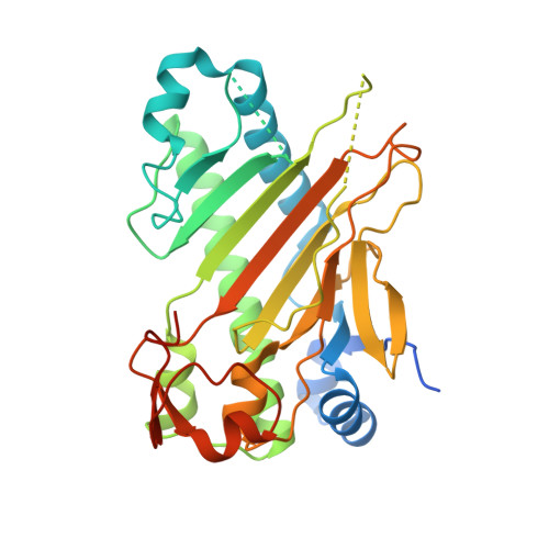
Conformational Flexibility of the C Terminus with Implications for Substrate Binding and Catalysis Revealed in a New Crystal Form of Deacetoxycephalosporin C Synthase
Oster, L.M., Terwisscha Van Scheltinga, A.C., Valegard, K., Mackenzie Hose, A., Dubus, A., Hajdu, J., Andersson, I.(2004) J Mol Biol 343: 157
- PubMed: 15381427
- DOI: https://doi.org/10.1016/j.jmb.2004.07.049
- Primary Citation of Related Structures:
1W28, 1W2A, 1W2N, 1W2O - PubMed Abstract:
Deacetoxycephalosporin C synthase (DAOCS) from Streptomyces clavuligerus catalyses the oxidative ring expansion of the penicillin nucleus into the nucleus of cephalosporins. The reaction requires dioxygen and 2-oxoglutarate as co-substrates to create a reactive iron-oxygen intermediate from a ferrous iron in the active site. The active enzyme is monomeric in solution. The structure of DAOCS was determined earlier from merohedrally twinned crystals where the last four C-terminal residues (308-311) of one molecule penetrate the active site of a neighbouring molecule, creating a cyclic trimeric structure in the crystal. Shortening the polypeptide chain from the C terminus by more than four residues diminishes activity. Here, we describe a new crystal form of DAOCS in which trimer formation is broken and the C-terminal arm is free. These crystals show no signs of twinning, and were obtained from DAOCS labelled with an N-terminal His-tag. The modified DAOCS is catalytically active. The free C-terminal arm protrudes into the solvent, and the C-terminal domain (residues 268-299) is rotated by about 16 degrees towards the active site. The last 12 residues (300-311) are disordered. Structures for various enzyme-substrate and enzyme-product complexes in the new crystal form confirm overlapping binding sites for penicillin and 2-oxoglutarate. The results support the notion that 2-oxoglutarate and dioxygen need to react first to produce an oxidizing iron species, followed by reaction with the penicillin substrate. The position of the penicillin nucleus is topologically similar in the two crystal forms, but the penicillin side-chain in the new non-twinned crystals overlaps with the position of residues 304-306 of the C-terminal arm in the twinned crystals. An analysis of the interactions between the C-terminal region and residues in the active site indicates that DAOCS could also accept polypeptide chains as ligands, and these could bind near the iron.
Organizational Affiliation:
Department of Molecular Biology, Swedish University of Agricultural Sciences, Box 590, S-75124 Uppsala.