1ST6

Crystal structure of a cytoskeletal protein


Experimental Data Snapshot

  • Method: X-RAY DIFFRACTION
  • Resolution: 3.10 Å
  • R-Value Free: 0.357 
  • R-Value Work: 0.316 
  • R-Value Observed: 0.316 

wwPDB Validation   3D Report Full Report


This is version 1.3 of the entry. See complete history


Literature

Structural basis for vinculin activation at sites of cell adhesion.

Bakolitsa, C.Cohen, D.M.Bankston, L.A.Bobkov, A.A.Cadwell, G.W.Jennings, L.Critchley, D.R.Craig, S.W.Liddington, R.C.

(2004) Nature 430: 583-586

  • DOI: https://doi.org/10.1038/nature02610
  • Primary Citation of Related Structures:  
    1ST6

  • PubMed Abstract: 

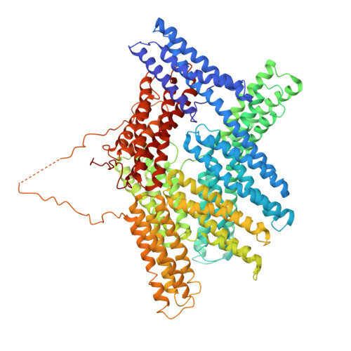
    Vinculin is a highly conserved intracellular protein with a crucial role in the maintenance and regulation of cell adhesion and migration. In the cytosol, vinculin adopts a default autoinhibited conformation. On recruitment to cell-cell and cell-matrix adherens-type junctions, vinculin becomes activated and mediates various protein-protein interactions that regulate the links between F-actin and the cadherin and integrin families of cell-adhesion molecules. Here we describe the crystal structure of the full-length vinculin molecule (1,066 amino acids), which shows a five-domain autoinhibited conformation in which the carboxy-terminal tail domain is held pincer-like by the vinculin head, and ligand binding is regulated both sterically and allosterically. We show that conformational changes in the head, tail and proline-rich domains are linked structurally and thermodynamically, and propose a combinatorial pathway to activation that ensures that vinculin is activated only at sites of cell adhesion when two or more of its binding partners are brought into apposition.


  • Organizational Affiliation

    Program on Cell Adhesion, The Burnham Institute, 10901 North Torrey Pines Road, La Jolla, California 92037, USA.


Macromolecules
Find similar proteins by:  (by identity cutoff)  |  3D Structure
Entity ID: 1
MoleculeChains Sequence LengthOrganismDetailsImage
Vinculin1,069Gallus gallusMutation(s): 0 
Gene Names: VCL
UniProt
Find proteins for P12003 (Gallus gallus)
Explore P12003 
Go to UniProtKB:  P12003
Entity Groups  
Sequence Clusters30% Identity50% Identity70% Identity90% Identity95% Identity100% Identity
UniProt GroupP12003
Sequence Annotations
Expand
  • Reference Sequence
Experimental Data & Validation

Experimental Data

  • Method: X-RAY DIFFRACTION
  • Resolution: 3.10 Å
  • R-Value Free: 0.357 
  • R-Value Work: 0.316 
  • R-Value Observed: 0.316 
  • Space Group: C 2 2 21
Unit Cell:
Length ( Å )Angle ( ˚ )
a = 56.02α = 90
b = 126.947β = 90
c = 351.87γ = 90
Software Package:
Software NamePurpose
CNSrefinement
DENZOdata reduction
XPREPdata reduction
SHELXDphasing
SHARPphasing

Structure Validation

View Full Validation Report


Entry History 

Deposition Data

Revision History  (Full details and data files)

  • Version 1.0: 2004-08-03
    Type: Initial release
  • Version 1.1: 2008-04-29
    Changes: Version format compliance
  • Version 1.2: 2011-07-13
    Changes: Version format compliance
  • Version 1.3: 2024-02-14
    Changes: Data collection, Database references