1QF5

DESIGN, SYNTHESIS, AND X-RAY CRYSTAL STRUCTURE OF AN ENZYME BOUND BISUBSTRATE HYBRID INHIBITOR OF ADENYLOSUCCINATE SYNTHETASE


Experimental Data Snapshot

  • Method: X-RAY DIFFRACTION
  • Resolution: 2.00 Å
  • R-Value Free: 0.228 
  • R-Value Work: 0.196 

wwPDB Validation   3D Report Full Report


This is version 1.4 of the entry. See complete history


Literature

An enzyme-bound bisubstrate hybrid inhibitor of adenylosuccinate synthetase

Hanessian, S.Lu, P.P.Sanceau, J.Y.Chemla, P.Gohda, K.Fonne-Pfister, R.Prade, L.Cowan-Jacob, S.W.

(1999) Angew Chem Int Ed Engl 38: 3159-3162

  • DOI: https://doi.org/10.1002/(sici)1521-3773(19991102)38:21<3159::aid-anie3159>3.0.co;2-2
  • Primary Citation of Related Structures:  
    1QF4, 1QF5

  • PubMed Abstract: 

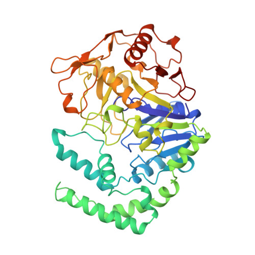
    Two relatively weak herbicides, hydantocidin phosphate and hadacidin were linked by a C(3) chain to afford a potent inhibitor (the 2S hybrid is shown) of the enzyme adenylosuccinate synthetase. The crystal structures of the bisubstrate-enzyme complexes were determined.


  • Organizational Affiliation

    Department of Chemistry, Université de Montréal, C.P. 6128, Succ. Centre-ville, Montreal, QC, H3C 3J7 (Canada).


Macromolecules
Find similar proteins by:  (by identity cutoff)  |  3D Structure
Entity ID: 1
MoleculeChains Sequence LengthOrganismDetailsImage
PROTEIN (ADENYLOSUCCINATE SYNTHETASE)431Escherichia coliMutation(s): 0 
EC: 6.3.4.4
UniProt
Find proteins for P0A7D4 (Escherichia coli (strain K12))
Explore P0A7D4 
Go to UniProtKB:  P0A7D4
Entity Groups  
Sequence Clusters30% Identity50% Identity70% Identity90% Identity95% Identity100% Identity
UniProt GroupP0A7D4
Sequence Annotations
Expand
  • Reference Sequence
Small Molecules
Ligands 4 Unique
IDChains Name / Formula / InChI Key2D Diagram3D Interactions
RPL
Query on RPL

Download Ideal Coordinates CCD File 
E [auth A](C8-S)-HYDANTOCIDIN 5'-PHOSPHATE
C13 H22 N3 O13 P
MAXSFYCTFIBEAR-FUIMDIGMSA-N
GDP
Query on GDP

Download Ideal Coordinates CCD File 
D [auth A]GUANOSINE-5'-DIPHOSPHATE
C10 H15 N5 O11 P2
QGWNDRXFNXRZMB-UUOKFMHZSA-N
PO4
Query on PO4

Download Ideal Coordinates CCD File 
B [auth A]PHOSPHATE ION
O4 P
NBIIXXVUZAFLBC-UHFFFAOYSA-K
MG
Query on MG

Download Ideal Coordinates CCD File 
C [auth A]MAGNESIUM ION
Mg
JLVVSXFLKOJNIY-UHFFFAOYSA-N
Binding Affinity Annotations 
IDSourceBinding Affinity
RPL PDBBind:  1QF5 IC50: 43 (nM) from 1 assay(s)
Binding MOAD:  1QF5 IC50: 43 (nM) from 1 assay(s)
Experimental Data & Validation

Experimental Data

  • Method: X-RAY DIFFRACTION
  • Resolution: 2.00 Å
  • R-Value Free: 0.228 
  • R-Value Work: 0.196 
  • Space Group: P 32 2 1
Unit Cell:
Length ( Å )Angle ( ˚ )
a = 81.75α = 90
b = 81.75β = 90
c = 158.7γ = 120
Software Package:
Software NamePurpose
AMoREphasing
X-PLORrefinement
MOSFLMdata reduction
CCP4data scaling

Structure Validation

View Full Validation Report


Entry History 

Deposition Data

Revision History  (Full details and data files)

  • Version 1.0: 1999-12-02
    Type: Initial release
  • Version 1.1: 2007-10-16
    Changes: Version format compliance
  • Version 1.2: 2011-07-13
    Changes: Derived calculations, Version format compliance
  • Version 1.3: 2019-11-20
    Changes: Database references
  • Version 1.4: 2023-08-16
    Changes: Data collection, Database references, Derived calculations, Refinement description