


Determination of the Structure of Papaya Protease Omega
Pickersgill, R.W., Rizkallah, P.J., Harris, G.W., Goodenough, P.W.(1991) Acta Crystallogr B 47: 766-771
Experimental Data Snapshot
wwPDB Validation 3D Report Full Report

(1991) Acta Crystallogr B 47: 766-771
Entity ID: 1 | |||||
|---|---|---|---|---|---|
| Molecule | Chains | Sequence Length | Organism | Details | Image |
| PROTEASE OMEGA | 216 | Carica papaya | Mutation(s): 0 EC: 3.4.22.30 | 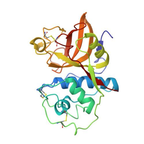 | |
UniProt | |||||
Find proteins for P10056 (Carica papaya) Explore P10056 Go to UniProtKB: P10056 | |||||
Entity Groups | |||||
| Sequence Clusters | 30% Identity50% Identity70% Identity90% Identity95% Identity100% Identity | ||||
| UniProt Group | P10056 | ||||
Sequence AnnotationsExpand | |||||
| |||||
| Ligands 1 Unique | |||||
|---|---|---|---|---|---|
| ID | Chains | Name / Formula / InChI Key | 2D Diagram | 3D Interactions | |
| HG Query on HG | B [auth A] | MERCURY (II) ION Hg BQPIGGFYSBELGY-UHFFFAOYSA-N |  | ||
| Length ( Å ) | Angle ( ˚ ) |
|---|---|
| a = 74.11 | α = 90 |
| b = 74.11 | β = 90 |
| c = 77.81 | γ = 120 |
| Software Name | Purpose |
|---|---|
| RESTRAIN | refinement |
RCSB PDB (citation) is hosted by
RCSB PDB is a member of the

