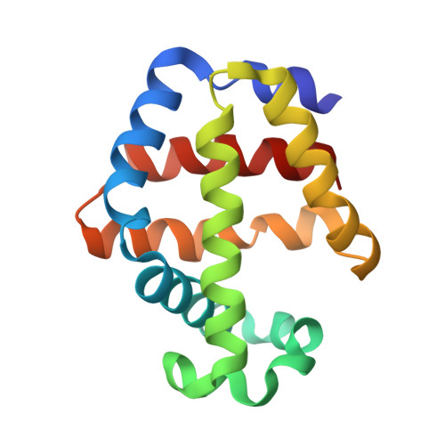
Ligand-linked structural transitions in crystals of a cooperative dimeric hemoglobin.
Knapp, J.E., Royer Jr., W.E.(2003) Biochemistry 42: 4640-4647
- PubMed: 12705827
- DOI: https://doi.org/10.1021/bi027136p
- Primary Citation of Related Structures:
1NWI, 1NWN, 1NXF - PubMed Abstract:
Cooperative ligand binding in the dimeric hemoglobin (HbI) from the blood clam Scapharca inaequivalvis is mediated primarily by tertiary structural changes, but with a small quaternary rearrangement (approximately 3 degrees), based on analysis of distinct crystal forms for ligated and unligated molecules. We report here ligand transition structures in both crystal forms. Binding CO to unligated HbI crystals results in a structure that approaches, but does not attain, the full allosteric transition. In contrast, removing CO from the HbI-CO crystals results in a structure that possesses all the key low affinity attributes previously identified from analysis of HbI crystals grown in the unligated state. Subsequent binding of CO shows the reversibility of this process. The observed structural changes include the quaternary rearrangement even under the constraints of lattice interactions, demonstrating that subunit rotation is an integral component of the ligand-linked structural transition in HbI. Analysis of both crystal forms, along with data from HbI mutants, suggests that the quaternary structural change is linked to the movement of the heme group, supporting a hypothesis that the heme movement is the central event that triggers cooperative ligand binding in this hemoglobin dimer. These results show both the effects of a crystal lattice in limiting quaternary structural transitions and provide the first example of complete allosteric transitions within another crystal lattice.
Organizational Affiliation:
Department of Biochemistry and Molecular Pharmacology, University of Massachusetts Medical School, Worcester, Massachusetts 01605, USA.