1N0I

Crystal Structure of Ferrochelatase with Cadmium bound at active site


Experimental Data Snapshot

  • Method: X-RAY DIFFRACTION
  • Resolution: 2.00 Å
  • R-Value Free: 0.273 
  • R-Value Work: 0.231 
  • R-Value Observed: 0.231 

wwPDB Validation   3D Report Full Report


This is version 1.4 of the entry. See complete history


Literature

Metal binding to Bacillus subtilis ferrochelatase and interaction between metal sites

Lecerof, D.Fodje, M.N.Leon, R.A.Olsson, U.Hansson, A.Sigfridsson, E.Ryde, U.Hansson, M.Al-Karadaghi, S.

(2003) J Biol Inorg Chem 8: 452-458

  • DOI: https://doi.org/10.1007/s00775-002-0436-1
  • Primary Citation of Related Structures:  
    1LD3, 1N0I

  • PubMed Abstract: 

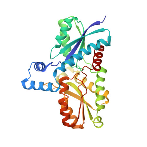
    Ferrochelatase, the terminal enzyme in heme biosynthesis, catalyses metal insertion into protoporphyrin IX. The location of the metal binding site with respect to the bound porphyrin substrate and the mode of metal binding are of central importance for understanding the mechanism of porphyrin metallation. In this work we demonstrate that Zn(2+), which is commonly used as substrate in assays of the ferrochelatase reaction, and Cd(2+), an inhibitor of the enzyme, bind to the invariant amino acids His183 and Glu264 and water molecules, all located within the porphyrin binding cleft. On the other hand, Mg(2+), which has been shown to bind close to the surface at 7 A from His183, was largely absent from its site. Activity measurements demonstrate that Mg(2+) has a stimulatory effect on the enzyme, lowering K(M) for Zn(2+) from 55 to 24 micro M. Changing one of the Mg(2+) binding residues, Glu272, to serine abolishes the effect of Mg(2+). It is proposed that prior to metal insertion the metal may form a sitting-atop (SAT) complex with the invariant His-Glu couple and the porphyrin. Metal binding to the Mg(2+) site may stimulate metal release from the protein ligands and its insertion into the porphyrin.


  • Organizational Affiliation

    Department of Molecular Biophysics, Center for Chemistry and Chemical Engineering, Lund University, PO Box 124, 221 00 Lund, Sweden.


Macromolecules
Find similar proteins by:  (by identity cutoff)  |  3D Structure
Entity ID: 1
MoleculeChains Sequence LengthOrganismDetailsImage
Ferrochelatase310Bacillus subtilisMutation(s): 0 
EC: 4.99.1.1
UniProt
Find proteins for P32396 (Bacillus subtilis (strain 168))
Explore P32396 
Go to UniProtKB:  P32396
Entity Groups  
Sequence Clusters30% Identity50% Identity70% Identity90% Identity95% Identity100% Identity
UniProt GroupP32396
Sequence Annotations
Expand
  • Reference Sequence
Experimental Data & Validation

Experimental Data

  • Method: X-RAY DIFFRACTION
  • Resolution: 2.00 Å
  • R-Value Free: 0.273 
  • R-Value Work: 0.231 
  • R-Value Observed: 0.231 
  • Space Group: P 21 21 21
Unit Cell:
Length ( Å )Angle ( ˚ )
a = 48.415α = 90
b = 49.878β = 90
c = 118.777γ = 90
Software Package:
Software NamePurpose
MAR345data collection
XDSdata reduction
CNSrefinement
XDSdata scaling
CNSphasing

Structure Validation

View Full Validation Report


Entry History 

Deposition Data

Revision History  (Full details and data files)

  • Version 1.0: 2003-05-27
    Type: Initial release
  • Version 1.1: 2008-04-28
    Changes: Version format compliance
  • Version 1.2: 2011-07-13
    Changes: Version format compliance
  • Version 1.3: 2017-10-11
    Changes: Data collection, Refinement description
  • Version 1.4: 2024-02-14
    Changes: Data collection, Database references, Derived calculations, Refinement description