1KZN

Crystal Structure of E. coli 24kDa Domain in Complex with Clorobiocin


Experimental Data Snapshot

  • Method: X-RAY DIFFRACTION
  • Resolution: 2.30 Å
  • R-Value Free: 0.267 
  • R-Value Work: 0.239 
  • R-Value Observed: 0.239 

wwPDB Validation   3D Report Full Report


Ligand Structure Quality Assessment 


This is version 1.3 of the entry. See complete history


Literature

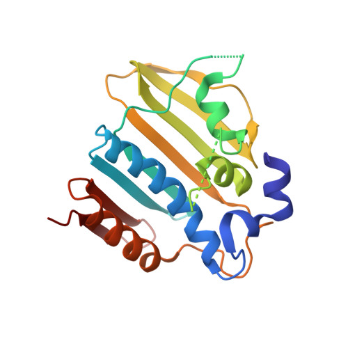
DNA gyrase interaction with coumarin-based inhibitors: the role of the hydroxybenzoate isopentenyl moiety and the 5'-methyl group of the noviose.

Lafitte, D.Lamour, V.Tsvetkov, P.O.Makarov, A.A.Klich, M.Deprez, P.Moras, D.Briand, C.Gilli, R.

(2002) Biochemistry 41: 7217-7223

  • DOI: https://doi.org/10.1021/bi0159837
  • Primary Citation of Related Structures:  
    1KZN

  • PubMed Abstract: 

    DNA gyrase is a major bacterial protein that is involved in replication and transcription and catalyzes the negative supercoiling of bacterial circular DNA. DNA gyrase is a known target for antibacterial agents since its blocking induces bacterial death. Quinolones, coumarins, and cyclothialidines have been designed to inhibit gyrase. Significant improvements can still be envisioned for a better coumarin-gyrase interaction. In this work, we obtained the crystal costructures of the natural coumarin clorobiocin and a synthetic analogue with the 24 kDa gyrase fragment. We used isothermal titration microcalorimetry and differential scanning calorimetry to obtain the thermodynamic parameters representative of the molecular interactions occurring during the binding process between coumarins and the 24 kDa gyrase fragment. We provide the first experimental evidence that clorobiocin binds gyrase with a stronger affinity than novobiocin. We also demonstrate the crucial role of both the hydroxybenzoate isopentenyl moiety and the 5'-alkyl group on the noviose of the coumarins in the binding affinity for gyrase.


  • Organizational Affiliation

    UMR CNRS 6032, UFR de Pharmacie, 27 Bd Jean Moulin, 13385 Marseille Cedex 5, France. daniel.lafitte@pharmacie.univ-mrs.fr


Macromolecules
Find similar proteins by:  (by identity cutoff)  |  3D Structure
Entity ID: 1
MoleculeChains Sequence LengthOrganismDetailsImage
DNA GYRASE SUBUNIT B205Escherichia coliMutation(s): 0 
EC: 5.99.1.3
UniProt
Find proteins for P0AES6 (Escherichia coli (strain K12))
Explore P0AES6 
Go to UniProtKB:  P0AES6
Entity Groups  
Sequence Clusters30% Identity50% Identity70% Identity90% Identity95% Identity100% Identity
UniProt GroupP0AES6
Sequence Annotations
Expand
  • Reference Sequence
Small Molecules
Ligands 1 Unique
IDChains Name / Formula / InChI Key2D Diagram3D Interactions
CBN
Query on CBN

Download Ideal Coordinates CCD File 
B [auth A]CLOROBIOCIN
C35 H37 Cl N2 O11
FJAQNRBDVKIIKK-LFLQOBSNSA-N
Binding Affinity Annotations 
IDSourceBinding Affinity
CBN BindingDB:  1KZN Kd: 1.2 (nM) from 1 assay(s)
IC50: 73 (nM) from 1 assay(s)
-TΔS: -1.14e+1 (kJ/mol) from 1 assay(s)
ΔH: -3.96e+1 (kJ/mol) from 1 assay(s)
ΔG: -5.10e+1 (kJ/mol) from 1 assay(s)
PDBBind:  1KZN Kd: 1.2 (nM) from 1 assay(s)
Binding MOAD:  1KZN Kd: 1.2 (nM) from 1 assay(s)
Experimental Data & Validation

Experimental Data

  • Method: X-RAY DIFFRACTION
  • Resolution: 2.30 Å
  • R-Value Free: 0.267 
  • R-Value Work: 0.239 
  • R-Value Observed: 0.239 
  • Space Group: P 21 21 21
Unit Cell:
Length ( Å )Angle ( ˚ )
a = 40.714α = 90
b = 47.629β = 90
c = 111.599γ = 90
Software Package:
Software NamePurpose
AMoREphasing
CNSrefinement
DENZOdata reduction
SCALEPACKdata scaling

Structure Validation

View Full Validation Report


Ligand Structure Quality Assessment 


Entry History 

Deposition Data

Revision History  (Full details and data files)

  • Version 1.0: 2002-06-19
    Type: Initial release
  • Version 1.1: 2008-04-28
    Changes: Version format compliance
  • Version 1.2: 2011-07-13
    Changes: Version format compliance
  • Version 1.3: 2024-02-14
    Changes: Data collection, Database references, Derived calculations