1IG3

Mouse Thiamin Pyrophosphokinase Complexed with Thiamin


Experimental Data Snapshot

  • Method: X-RAY DIFFRACTION
  • Resolution: 1.90 Å
  • R-Value Free: 0.239 
  • R-Value Work: 0.214 
  • R-Value Observed: 0.214 

wwPDB Validation   3D Report Full Report


Ligand Structure Quality Assessment 


This is version 1.3 of the entry. See complete history


Literature

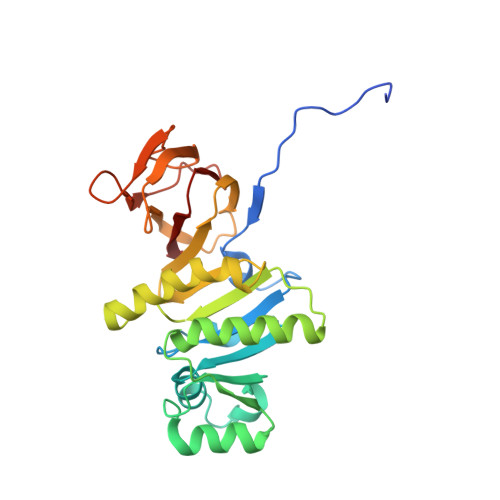
Crystal structure of thiamin pyrophosphokinase.

Timm, D.E.Liu, J.Baker, L.J.Harris, R.A.

(2001) J Mol Biol 310: 195-204

  • DOI: https://doi.org/10.1006/jmbi.2001.4727
  • Primary Citation of Related Structures:  
    1IG3

  • PubMed Abstract: 

    Thiamin pyrophosphate (TPP) is a coenzyme derived from vitamin B1 (thiamin). TPP synthesis in eukaryotes requires thiamin pyrophosphokinase (TPK), which catalyzes the transfer of a pyrophosphate group from ATP to thiamin. TPP is essential for central metabolic processes, including the formation of acetyl CoA from glucose and the Krebs cycle. Deficiencies in human thiamin metabolism result in beriberi and Wernicke encephalopathy. The crystal structure of mouse TPK was determined by multiwavelength anomalous diffraction at 2.4 A resolution, and the structure of TPK complexed with thiamin has been refined at 1.9 A resolution. The TPK polypeptide folds as an alpha/beta-domain and a beta-sandwich domain, which share a central ten-stranded mixed beta-sheet. TPK subunits associate as a dimer, and thiamin is bound in the dimer interface. Despite lacking apparent sequence homology with other proteins, the alpha/beta-domain resembles the Rossman fold and is similar to other kinase structures, including another pyrophosphokinase and a thiamin biosynthetic enzyme. Comparison of mouse and yeast TPK structures reveals differences that could be exploited in developing species-specific inhibitors of potential use as antimicrobial agents.


  • Organizational Affiliation

    Department of Biochemistry and Molecular Biology, Indiana University School of Medicine, Indianapolis, IN 46202, USA. dtimm@iupui.edu


Macromolecules
Find similar proteins by:  (by identity cutoff)  |  3D Structure
Entity ID: 1
MoleculeChains Sequence LengthOrganismDetailsImage
thiamin pyrophosphokinase
A, B
263Mus musculusMutation(s): 0 
EC: 2.7.6.2
UniProt & NIH Common Fund Data Resources
Find proteins for Q9R0M5 (Mus musculus)
Explore Q9R0M5 
Go to UniProtKB:  Q9R0M5
IMPC:  MGI:1352500
Entity Groups  
Sequence Clusters30% Identity50% Identity70% Identity90% Identity95% Identity100% Identity
UniProt GroupQ9R0M5
Sequence Annotations
Expand
  • Reference Sequence
Small Molecules
Ligands 2 Unique
IDChains Name / Formula / InChI Key2D Diagram3D Interactions
VIB
Query on VIB

Download Ideal Coordinates CCD File 
L [auth A],
R [auth B]
3-(4-AMINO-2-METHYL-PYRIMIDIN-5-YLMETHYL)-5-(2-HYDROXY-ETHYL)-4-METHYL-THIAZOL-3-IUM
C12 H17 N4 O S
JZRWCGZRTZMZEH-UHFFFAOYSA-N
SO4
Query on SO4

Download Ideal Coordinates CCD File 
C [auth A]
D [auth A]
E [auth A]
F [auth A]
G [auth A]
C [auth A],
D [auth A],
E [auth A],
F [auth A],
G [auth A],
H [auth A],
I [auth A],
J [auth A],
K [auth A],
M [auth B],
N [auth B],
O [auth B],
P [auth B],
Q [auth B]
SULFATE ION
O4 S
QAOWNCQODCNURD-UHFFFAOYSA-L
Experimental Data & Validation

Experimental Data

  • Method: X-RAY DIFFRACTION
  • Resolution: 1.90 Å
  • R-Value Free: 0.239 
  • R-Value Work: 0.214 
  • R-Value Observed: 0.214 
  • Space Group: P 31 2 1
Unit Cell:
Length ( Å )Angle ( ˚ )
a = 89.8α = 90
b = 89.8β = 90
c = 141.1γ = 120
Software Package:
Software NamePurpose
CNSrefinement
DENZOdata reduction
SCALEPACKdata scaling
CNSphasing

Structure Validation

View Full Validation Report


Ligand Structure Quality Assessment 


Entry History 

Deposition Data

Revision History  (Full details and data files)

  • Version 1.0: 2001-04-25
    Type: Initial release
  • Version 1.1: 2008-04-27
    Changes: Version format compliance
  • Version 1.2: 2011-07-13
    Changes: Version format compliance
  • Version 1.3: 2024-02-07
    Changes: Data collection, Database references, Derived calculations, Structure summary