Cooperative zinc binding in a staphylococcal enterotoxin A mutant mimics the SEA-MHC class II interaction
Hakansson, M., Antonsson, P., Bjork, P., Svensson, L.A.(2001) J Biol Inorg Chem 6: 757-762
- PubMed: 11713682
- DOI: https://doi.org/10.1007/s007750100251
- Primary Citation of Related Structures:
1I4G, 1I4H - PubMed Abstract:
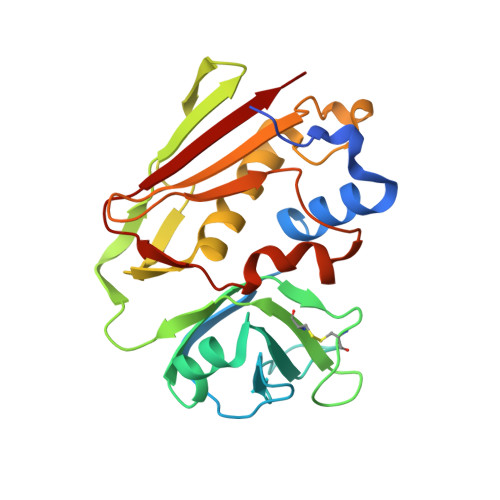
The structure of a mutant form of staphylococcal enterotoxin A (SEA) has been determined to 2.1 A resolution. The studied SEA substitution H187-->A187 (SEAH187A) leads to an almost 10-fold reduction of the binding to major histocompatibility complex (MHC) class II. H187 is important for this interaction since it coordinates Zn2+. The zinc ion is thought to hold MHC class II and SEA together in a complex. Interestingly, only one of two molecules in the asymmetric unit binds Zn2+. H225, D227, a water molecule, and H44 from a symmetry-related molecule ligate Zn2+. The symmetry-related histidine is necessary for this substituted Zn2+ site to bind to Zn2+ at low zinc concentration (no Zn2+ added). Since a water molecule replaces the missing H187, H44 binds Zn2+ at the position where betaH81 from MHC class II probably will bind. Dynamic light scattering analysis reveals that in solution as well as in the crystal lattice the SEA(H187A) mutant forms aggregates. The substitution per se does not cause aggregation since wild-type SEA also forms aggregates. Addition of EDTA reduces the size of the aggregates, indicating a cross-linking function of Zn2+. In agreement with the biological function, the aggregation is weak (i.e. not revealed by gel filtration) and non-specific.
Organizational Affiliation:
Department of Molecular Biophysics, Center for Chemistry and Chemical Engineering, P.O. Box 124, 221 00 Lund, Sweden. maria.hakansson@mbfys.lu.se