1I1C

NON-FCRN BINDING FC FRAGMENT OF RAT IGG2A


Experimental Data Snapshot

  • Method: X-RAY DIFFRACTION
  • Resolution: 2.70 Å
  • R-Value Free: 0.279 
  • R-Value Work: 0.241 

wwPDB Validation   3D Report Full Report


This is version 2.2 of the entry. See complete history


Literature

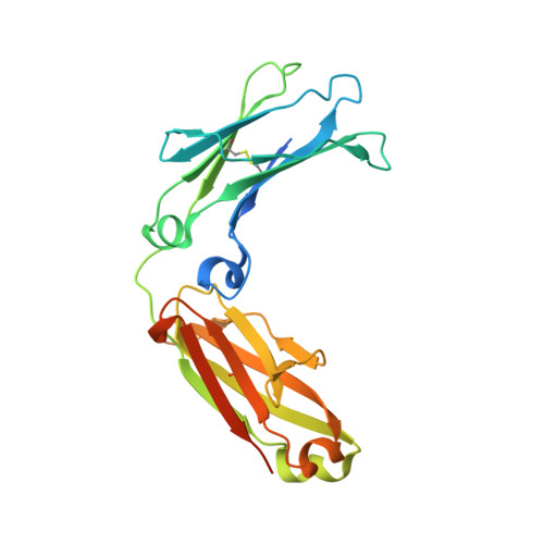
Crystal structure at 2.8 A of an FcRn/heterodimeric Fc complex: mechanism of pH-dependent binding.

Martin, W.L.West Jr., A.P.Gan, L.Bjorkman, P.J.

(2001) Mol Cell 7: 867-877

  • DOI: https://doi.org/10.1016/s1097-2765(01)00230-1
  • Primary Citation of Related Structures:  
    1I1A, 1I1C

  • PubMed Abstract: 

    The neonatal Fc receptor (FcRn) transports immunoglobulin G (IgG) across epithelia, binding IgG in acidic vesicles (pH < or = 6.5) and releasing IgG in the blood at pH 7.4. Well-ordered FcRn/Fc crystals are prevented by the formation of "oligomeric ribbons" of FcRn dimers bridged by Fc homodimers, thus we crystallized a 1:1 complex between rat FcRn and a heterodimeric Fc containing only one FcRn binding site. The 2.8 A complex structure demonstrates that FcRn uses its alpha2 and beta2-microglobulin domains and carbohydrate to interact with the Fc C(gamma)2-C(gamma)3 interface. The structure reveals conformational changes in Fc and three titratable salt bridges that confer pH-dependent binding, and can be used to guide rational design of therapeutic IgGs with longer serum half-lives.


  • Organizational Affiliation

    Division of Biology, California Institute of Technology, Pasadena, CA 91125, USA.


Macromolecules
Find similar proteins by:  (by identity cutoff)  |  3D Structure
Entity ID: 1
MoleculeChains Sequence LengthOrganismDetailsImage
IG GAMMA-2A CHAIN C REGION
A, B
239Rattus norvegicusMutation(s): 6 
UniProt
Find proteins for P20760 (Rattus norvegicus)
Explore P20760 
Go to UniProtKB:  P20760
Entity Groups  
Sequence Clusters30% Identity50% Identity70% Identity90% Identity95% Identity100% Identity
UniProt GroupP20760
Sequence Annotations
Expand
  • Reference Sequence
Oligosaccharides

Help

Entity ID: 2
MoleculeChains Length2D Diagram Glycosylation3D Interactions
2-acetamido-2-deoxy-beta-D-glucopyranose-(1-2)-alpha-D-mannopyranose-(1-3)-[2-acetamido-2-deoxy-beta-D-glucopyranose-(1-2)-alpha-D-mannopyranose-(1-6)]beta-D-mannopyranose-(1-4)-2-acetamido-2-deoxy-beta-D-glucopyranose-(1-4)-[beta-L-fucopyranose-(1-6)]2-acetamido-2-deoxy-beta-D-glucopyranose
C
8N-Glycosylation
Glycosylation Resources
GlyTouCan:  G59451NL
GlyCosmos:  G59451NL
GlyGen:  G59451NL
Entity ID: 3
MoleculeChains Length2D Diagram Glycosylation3D Interactions
2-acetamido-2-deoxy-beta-D-glucopyranose-(1-2)-alpha-D-mannopyranose-(1-3)-[2-acetamido-2-deoxy-beta-D-glucopyranose-(1-2)-alpha-D-mannopyranose-(1-6)]beta-D-mannopyranose-(1-4)-2-acetamido-2-deoxy-beta-D-glucopyranose-(1-4)-[alpha-L-fucopyranose-(1-6)]2-acetamido-2-deoxy-beta-D-glucopyranose
D
8N-Glycosylation
Glycosylation Resources
GlyTouCan:  G80858MF
GlyCosmos:  G80858MF
GlyGen:  G80858MF
Experimental Data & Validation

Experimental Data

  • Method: X-RAY DIFFRACTION
  • Resolution: 2.70 Å
  • R-Value Free: 0.279 
  • R-Value Work: 0.241 
  • Space Group: P 1 21 1
Unit Cell:
Length ( Å )Angle ( ˚ )
a = 42.503α = 90
b = 73.362β = 102.74
c = 82.157γ = 90
Software Package:
Software NamePurpose
DENZOdata reduction
SCALEPACKdata scaling
AMoREphasing
CNSrefinement

Structure Validation

View Full Validation Report


Entry History 

Deposition Data

Revision History  (Full details and data files)

  • Version 1.0: 2001-02-14
    Type: Initial release
  • Version 1.1: 2008-04-27
    Changes: Version format compliance
  • Version 1.2: 2011-07-13
    Changes: Non-polymer description, Version format compliance
  • Version 2.0: 2020-07-29
    Type: Remediation
    Reason: Carbohydrate remediation
    Changes: Advisory, Atomic model, Data collection, Database references, Derived calculations, Structure summary
  • Version 2.1: 2021-10-27
    Changes: Database references, Structure summary
  • Version 2.2: 2023-08-09
    Changes: Data collection, Refinement description