1G17

CRYSTAL STRUCTURE OF SEC4-GUANOSINE-5'-(BETA,GAMMA)-IMIDOTRIPHOSPHATE


Experimental Data Snapshot

  • Method: X-RAY DIFFRACTION
  • Resolution: 2.00 Å
  • R-Value Free: 0.292 
  • R-Value Work: 0.264 
  • R-Value Observed: 0.264 

wwPDB Validation   3D Report Full Report


Ligand Structure Quality Assessment 


This is version 1.3 of the entry. See complete history


Literature

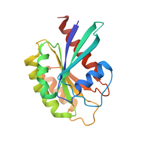
Crystal structures of a Rab protein in its inactive and active conformations.

Stroupe, C.Brunger, A.T.

(2000) J Mol Biol 304: 585-598

  • DOI: https://doi.org/10.1006/jmbi.2000.4236
  • Primary Citation of Related Structures:  
    1G16, 1G17

  • PubMed Abstract: 

    We have determined crystal structures of Sec4, a member of the Rab family in the G protein superfamily, in two states: bound to GDP, and to a non-hydrolyzable GTP analog, guanosine-5'-(beta, gamma)-imidotriphosphate (GppNHp). This represents the first structure of a Rab protein bound to GDP. Sec4 in both states grossly resembles other G proteins bound to GDP and GppNHp. In Sec4-GppNHp, structural features common to active Rab proteins are observed. In Sec4-GDP, the switch I region is highly disordered and displaced relative to the switch I region of Ras-GDP. In two of the four molecules of Sec4-GDP in the asymmetric unit of the Sec4-GDP crystals, the switch II region adopts a conformation similar to that seen in the structure of the small G protein Ran bound to GDP. This allows residues threonine 76, glutamate 80, and arginine 81 of Sec4 to make contacts with other conserved residues and water molecules important for nucleotide binding. In the other two molecules in the asymmetric unit, these interactions do not take place. This structural variability in both the switch I and switch II regions of GDP-bound Sec4 provides a possible explanation for the high off-rate of GDP bound to Sec4, and suggests a mechanism for regulation of the GTPase cycle of Rab proteins by GDI proteins.


  • Organizational Affiliation

    The Howard Hughes Medical Institute and Departments of Molecular and Cellular Physiology, Stanford University, Stanford, CA, 94305-548, USA.


Macromolecules
Find similar proteins by:  (by identity cutoff)  |  3D Structure
Entity ID: 1
MoleculeChains Sequence LengthOrganismDetailsImage
RAS-RELATED PROTEIN SEC4
A, B
170Saccharomyces cerevisiaeMutation(s): 0 
UniProt
Find proteins for P07560 (Saccharomyces cerevisiae (strain ATCC 204508 / S288c))
Explore P07560 
Go to UniProtKB:  P07560
Entity Groups  
Sequence Clusters30% Identity50% Identity70% Identity90% Identity95% Identity100% Identity
UniProt GroupP07560
Sequence Annotations
Expand
  • Reference Sequence
Experimental Data & Validation

Experimental Data

  • Method: X-RAY DIFFRACTION
  • Resolution: 2.00 Å
  • R-Value Free: 0.292 
  • R-Value Work: 0.264 
  • R-Value Observed: 0.264 
  • Space Group: P 65
Unit Cell:
Length ( Å )Angle ( ˚ )
a = 84.504α = 90
b = 84.504β = 90
c = 86.469γ = 120
Software Package:
Software NamePurpose
DENZOdata reduction
SCALEPACKdata scaling
CNSrefinement
CNSphasing

Structure Validation

View Full Validation Report


Ligand Structure Quality Assessment 


Entry History 

Deposition Data

Revision History  (Full details and data files)

  • Version 1.0: 2000-12-11
    Type: Initial release
  • Version 1.1: 2008-04-27
    Changes: Version format compliance
  • Version 1.2: 2011-07-13
    Changes: Version format compliance
  • Version 1.3: 2024-02-07
    Changes: Data collection, Database references, Derived calculations