1FA9

HUMAN LIVER GLYCOGEN PHOSPHORYLASE A COMPLEXED WITH AMP


Experimental Data Snapshot

  • Method: X-RAY DIFFRACTION
  • Resolution: 2.40 Å
  • R-Value Free: 0.295 
  • R-Value Work: 0.235 
  • R-Value Observed: 0.221 

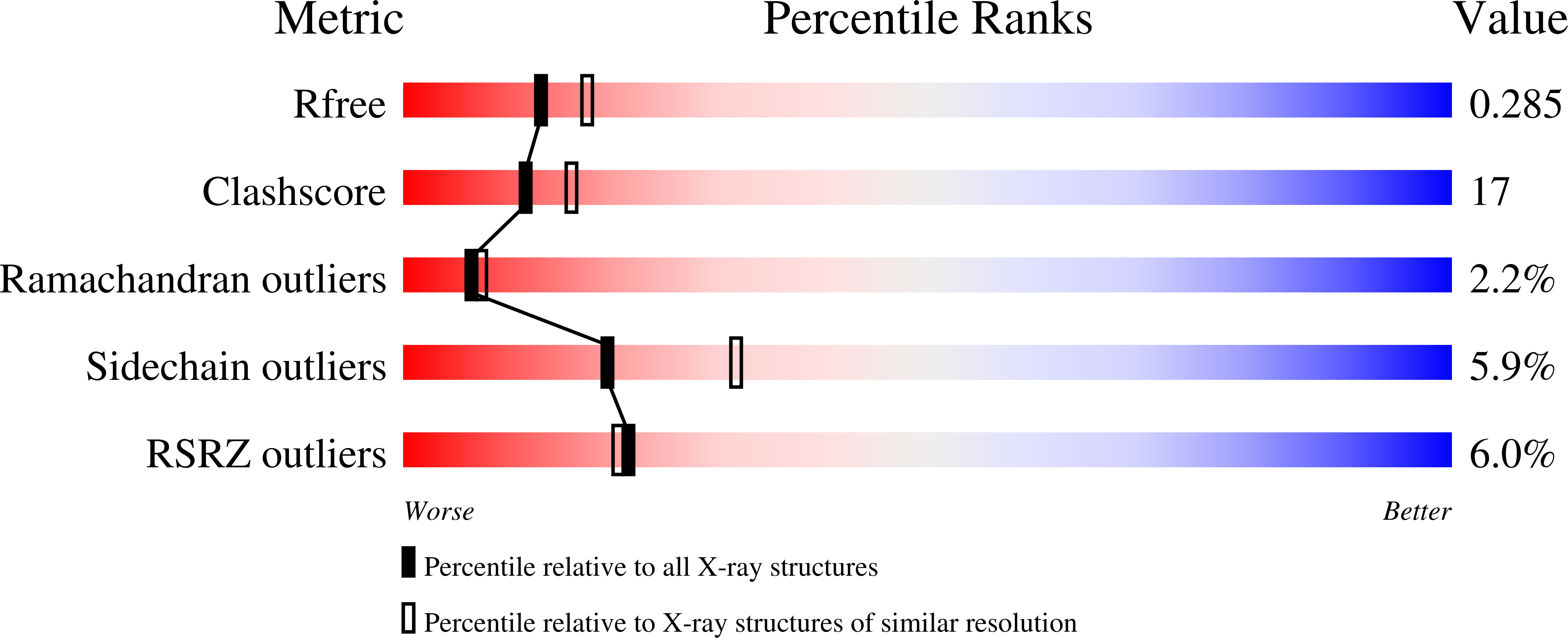
wwPDB Validation   3D Report Full Report


Ligand Structure Quality Assessment 


This is version 1.5 of the entry. See complete history


Literature

Activation of human liver glycogen phosphorylase by alteration of the secondary structure and packing of the catalytic core.

Rath, V.L.Ammirati, M.LeMotte, P.K.Fennell, K.F.Mansour, M.N.Danley, D.E.Hynes, T.R.Schulte, G.K.Wasilko, D.J.Pandit, J.

(2000) Mol Cell 6: 139-148

  • Primary Citation of Related Structures:  
    1FA9, 1FC0

  • PubMed Abstract: 

    Glycogen phosphorylases catalyze the breakdown of glycogen to glucose-1-phosphate, which enters glycolysis to fulfill the energetic requirements of the organism. Maintaining control of blood glucose levels is critical in minimizing the debilitating effects of diabetes, making liver glycogen phosphorylase a potential therapeutic target. To support inhibitor design, we determined the crystal structures of the active and inactive forms of human liver glycogen phosphorylase a. During activation, forty residues of the catalytic site undergo order/disorder transitions, changes in secondary structure, or packing to reorganize the catalytic site for substrate binding and catalysis. Knowing the inactive and active conformations of the liver enzyme and how each differs from its counterpart in muscle phosphorylase provides the basis for designing inhibitors that bind preferentially to the inactive conformation of the liver isozyme.


  • Organizational Affiliation

    Exploratory Medicinal Sciences, Global Research and Development, Groton Laboratories, Pfizer, Inc, Connecticut 06340, USA.


Macromolecules
Find similar proteins by:  (by identity cutoff)  |  3D Structure
Entity ID: 1
MoleculeChains Sequence LengthOrganismDetailsImage
GLYCOGEN PHOSPHORYLASE, LIVER FORM846Homo sapiensMutation(s): 1 
EC: 2.4.1.1
UniProt & NIH Common Fund Data Resources
Find proteins for P06737 (Homo sapiens)
Explore P06737 
Go to UniProtKB:  P06737
PHAROS:  P06737
GTEx:  ENSG00000100504 
Entity Groups  
Sequence Clusters30% Identity50% Identity70% Identity90% Identity95% Identity100% Identity
UniProt GroupP06737
Sequence Annotations
Expand
  • Reference Sequence
Small Molecules
Modified Residues  1 Unique
IDChains TypeFormula2D DiagramParent
SEP
Query on SEP
A
L-PEPTIDE LINKINGC3 H8 N O6 PSER
Experimental Data & Validation

Experimental Data

  • Method: X-RAY DIFFRACTION
  • Resolution: 2.40 Å
  • R-Value Free: 0.295 
  • R-Value Work: 0.235 
  • R-Value Observed: 0.221 
  • Space Group: P 31 2 1
Unit Cell:
Length ( Å )Angle ( ˚ )
a = 123.91α = 90
b = 123.91β = 90
c = 127.68γ = 120
Software Package:
Software NamePurpose
X-PLORmodel building
X-PLORrefinement
DENZOdata reduction
SCALEPACKdata scaling
X-PLORphasing

Structure Validation

View Full Validation Report


Ligand Structure Quality Assessment 


Entry History 

Deposition Data

Revision History  (Full details and data files)

  • Version 1.0: 2000-08-25
    Type: Initial release
  • Version 1.1: 2008-04-27
    Changes: Version format compliance
  • Version 1.2: 2011-07-13
    Changes: Derived calculations, Version format compliance
  • Version 1.3: 2018-01-31
    Changes: Experimental preparation
  • Version 1.4: 2020-07-29
    Type: Remediation
    Reason: Carbohydrate remediation
    Changes: Data collection, Database references, Derived calculations, Structure summary
  • Version 1.5: 2023-08-09
    Changes: Data collection, Database references, Refinement description, Structure summary