Structural basis for uracil DNA glycosylase interaction with uracil: NMR study.
Ghosh, M., Vinay Kumar, N., Varshney, U., Chary, K.V.(2000) Nucleic Acids Res 28: 1906-1912
- PubMed: 10756190
- DOI: https://doi.org/10.1093/nar/28.9.1906
- Primary Citation of Related Structures:
1DGO - PubMed Abstract:
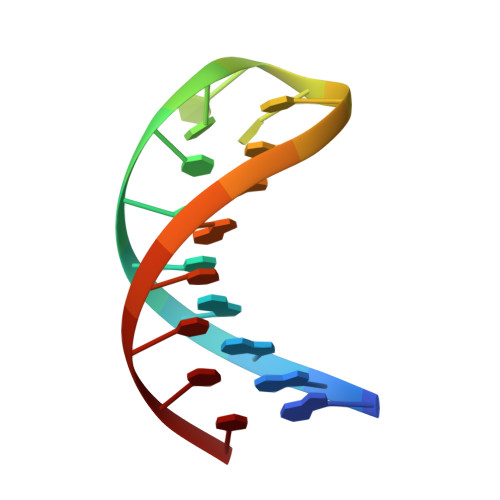
Two dimensional (2D) NMR and molecular dynamics simulations have been used to determine the three dimensional (3D) structure of a hairpin DNA, d-CTA-GAGGATCC-TUTT-GGATCCT (22mer; abbreviated as U2-hairpin), which has uracil at the second position from the 5' end of the tetraloop. The(1)H resonances of this hairpin have been assigned almost completely. NMR restrained molecular dynamics and energy minimization procedures have been used to describe the 3D structure of U2-hairpin. This study establishes that the stem of the hairpin adopts a right-handed B-DNA conformation, while the T(12)and T(15)nucleotides stack upon 3' and 5' ends of the stem, respectively. Further, T(14)stacks upon both T(12)and T(15). Though U(13)partially stacks upon T(14), no stacking interaction is observed between U(13)and T(12). All the individual nucleotide bases belonging to the stem and T(12)and T(15)of the loop adopt ' anti ' conformation with respect to their sugar moiety, while the U(13)and T(14)of the loop are in ' syn ' conformation. The turning phosphate in the loop is located between T(13)and T(14). This study and a concurrent NMR structural study on yet another hairpin DNA d-CTAGAGGAATAA-TTTU-GGATCCT (22mer; abbreviated as U4-hairpin), with uracil at the fourth position from the 5' end of the tetraloop throw light upon various interactions which have been reported between Escherichia coli uracil DNA glycosylase (UDG) and uracil containing DNA. The epsilon of T(12)and alpha, beta, gamma, epsilon and zeta of U(13)and gamma of T(14), which partially influence the local conformation of U(13)in U2-hairpin are all locked in ' trans ' conformation. Such stretched out backbone conformation in the vicinity of U(13)could be the reason as to why the U2-hairpin is found to be the poor substrate for its interaction with UDG compared to the other substrates in which the uracil is at first, third and fourth positions of the tetraloop from its 5' end, as reported earlier by Vinay and Varshney. This study shows that UDG actively promotes the flipping of uracil from a stacked conformation and rules out the possibility of UDG recognizing the flipped out uracil bases.
Organizational Affiliation:
Department of Chemical Sciences, Tata Institute of Fundamental Research, Homi Bhabha Road, Colaba, Mumbai 400 005, India.