1AP2

SINGLE CHAIN FV OF C219


Experimental Data Snapshot

  • Method: X-RAY DIFFRACTION
  • Resolution: 2.36 Å
  • R-Value Free: 0.284 
  • R-Value Work: 0.204 
  • R-Value Observed: 0.204 

wwPDB Validation   3D Report Full Report


This is version 1.3 of the entry. See complete history


Literature

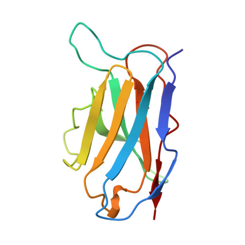
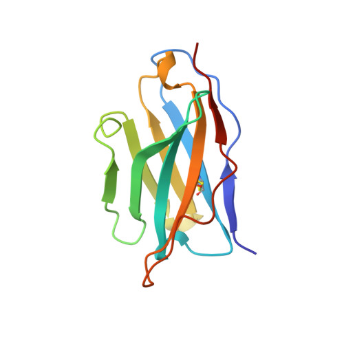
A single chain Fv fragment of P-glycoprotein-specific monoclonal antibody C219. Design, expression, and crystal structure at 2.4 A resolution.

Hoedemaeker, F.J.Signorelli, T.Johns, K.Kuntz, D.A.Rose, D.R.

(1997) J Biol Chem 272: 29784-29789

  • DOI: https://doi.org/10.1074/jbc.272.47.29784
  • Primary Citation of Related Structures:  
    1AP2

  • PubMed Abstract: 

    A construct encoding a single chain variable fragment of the anti-P-glycoprotein monoclonal antibody C219 was made by combining the coding sequences for the heavy and light chain variable domains with a sequence encoding the flexible linker (GGGGS)3, an OmpA signal sequence, a c-myc identification tag, and a five-histidine purification tag. The construct was expressed in Escherichia coli and purified from the periplasmic fraction using a nickel chelate column and ion exchange chromatography. Three-step Western blot analysis showed that the construct retains binding affinity for P-glycoprotein. Crystals of 1.0 x 0.2 x 0.2 mm were grown in 100 mM citrate, pH 4.5, 21% polyethylene glycol 6000 in the presence of low concentrations of subtilisin, resulting in proteolytic removal of the linker and purification tags. The structure was solved to a resolution of 2.4 A with an R factor of 20.6, an Rfree of 28.5, and good stereochemistry. This result could lead to a clinically useful product based on antibody C219 for the diagnosis of P-glycoprotein-mediated multidrug resistance. The molecule will also be useful in biophysical studies of functional domains of P-glycoprotein, as well as studies of the intact molecule.


  • Organizational Affiliation

    Ontario Cancer Institute and Department of Medical Biophysics, University of Toronto, Toronto M5G 2M9, Ontario, Canada.


Macromolecules
Find similar proteins by:  (by identity cutoff)  |  3D Structure
Entity ID: 1
MoleculeChains Sequence LengthOrganismDetailsImage
MONOCLONAL ANTIBODY C219
A, C
113Mus musculusMutation(s): 0 
Gene Names: CDNA
Entity Groups  
Sequence Clusters30% Identity50% Identity70% Identity90% Identity95% Identity100% Identity
Sequence Annotations
Expand
  • Reference Sequence
Find similar proteins by:  (by identity cutoff)  |  3D Structure
Entity ID: 2
MoleculeChains Sequence LengthOrganismDetailsImage
MONOCLONAL ANTIBODY C219
B, D
123Mus musculusMutation(s): 0 
Gene Names: CDNA
Entity Groups  
Sequence Clusters30% Identity50% Identity70% Identity90% Identity95% Identity100% Identity
Sequence Annotations
Expand
  • Reference Sequence
Experimental Data & Validation

Experimental Data

  • Method: X-RAY DIFFRACTION
  • Resolution: 2.36 Å
  • R-Value Free: 0.284 
  • R-Value Work: 0.204 
  • R-Value Observed: 0.204 
  • Space Group: P 21 21 21
Unit Cell:
Length ( Å )Angle ( ˚ )
a = 59.04α = 90
b = 64.35β = 90
c = 154.11γ = 90
Software Package:
Software NamePurpose
SDMSdata collection
SDMSdata reduction
X-PLORmodel building
X-PLORrefinement
SDMSdata scaling
X-PLORphasing

Structure Validation

View Full Validation Report


Entry History 

Deposition Data

Revision History  (Full details and data files)

  • Version 1.0: 1997-12-24
    Type: Initial release
  • Version 1.1: 2008-03-24
    Changes: Version format compliance
  • Version 1.2: 2011-07-13
    Changes: Version format compliance
  • Version 1.3: 2023-08-02
    Changes: Database references, Other, Refinement description