7YWP

Closed conformation of Oligopeptidase B from Serratia proteomaculans with covalently bound TCK


Experimental Data Snapshot

  • Method: X-RAY DIFFRACTION
  • Resolution: 2.20 Å
  • R-Value Free: 0.254 
  • R-Value Work: 0.184 
  • R-Value Observed: 0.188 

wwPDB Validation   3D Report Full Report


Ligand Structure Quality Assessment 


This is version 1.2 of the entry. See complete history


Literature

Crystal Structure of Inhibitor-Bound Bacterial Oligopeptidase B in the Closed State: Similarity and Difference between Protozoan and Bacterial Enzymes.

Petrenko, D.E.Karlinsky, D.M.Gordeeva, V.D.Arapidi, G.P.Britikova, E.V.Britikov, V.V.Nikolaeva, A.Y.Boyko, K.M.Timofeev, V.I.Kuranova, I.P.Mikhailova, A.G.Bocharov, E.V.Rakitina, T.V.

(2023) Int J Mol Sci 24

  • DOI: https://doi.org/10.3390/ijms24032286
  • Primary Citation of Related Structures:  
    7YWP

  • PubMed Abstract: 

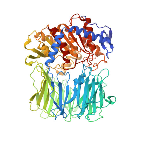
    The crystal structure of bacterial oligopeptidase B from Serratia proteamaculans (SpOpB) in complex with a chloromethyl ketone inhibitor was determined at 2.2 Å resolution. SpOpB was crystallized in a closed (catalytically active) conformation. A single inhibitor molecule bound simultaneously to the catalytic residues S532 and H652 mimicked a tetrahedral intermediate of the catalytic reaction. A comparative analysis of the obtained structure and the structure of OpB from Trypanosoma brucei (TbOpB) in a closed conformation showed that in both enzymes, the stabilization of the D-loop (carrying the catalytic D) in a position favorable for the formation of a tetrahedral complex occurs due to interaction with the neighboring loop from the β-propeller. However, the modes of interdomain interactions were significantly different for bacterial and protozoan OpBs. Instead of a salt bridge (as in TbOpB), in SpOpB, a pair of polar residues following the catalytic D617 and a pair of neighboring arginine residues from the β-propeller domain formed complementary oppositely charged surfaces. Bioinformatics analysis and structural modeling show that all bacterial OpBs can be divided into two large groups according to these two modes of D-loop stabilization in closed conformations.


  • Organizational Affiliation

    National Research Center "Kurchatov Institute", 123182 Moscow, Russia.


Macromolecules
Find similar proteins by:  (by identity cutoff)  |  3D Structure
Entity ID: 1
MoleculeChains Sequence LengthOrganismDetailsImage
Oligopeptidase B676Serratia proteamaculansMutation(s): 0 
Gene Names: opdB
EC: 3.4.21.83
UniProt
Find proteins for B3VI58 (Serratia proteamaculans)
Explore B3VI58 
Go to UniProtKB:  B3VI58
Entity Groups  
Sequence Clusters30% Identity50% Identity70% Identity90% Identity95% Identity100% Identity
UniProt GroupB3VI58
Sequence Annotations
Expand
  • Reference Sequence
Small Molecules
Ligands 1 Unique
IDChains Name / Formula / InChI Key2D Diagram3D Interactions
TCK
Query on TCK

Download Ideal Coordinates CCD File 
B [auth A]N-[(1S)-5-amino-1-(chloroacetyl)pentyl]-4-methylbenzenesulfonamide
C14 H21 Cl N2 O3 S
RDFCSSHDJSZMTQ-ZDUSSCGKSA-N
Biologically Interesting Molecules (External Reference) 1 Unique
Experimental Data & Validation

Experimental Data

  • Method: X-RAY DIFFRACTION
  • Resolution: 2.20 Å
  • R-Value Free: 0.254 
  • R-Value Work: 0.184 
  • R-Value Observed: 0.188 
  • Space Group: P 21 21 21
Unit Cell:
Length ( Å )Angle ( ˚ )
a = 75.529α = 90
b = 89.66β = 90
c = 108.65γ = 90
Software Package:
Software NamePurpose
REFMACrefinement
PDB_EXTRACTdata extraction
iMOSFLMdata reduction
SCALAdata scaling
PHASERphasing

Structure Validation

View Full Validation Report


Ligand Structure Quality Assessment 


Entry History & Funding Information

Deposition Data


Funding OrganizationLocationGrant Number
Not funded--

Revision History  (Full details and data files)

  • Version 1.0: 2023-02-22
    Type: Initial release
  • Version 1.1: 2023-03-01
    Changes: Database references
  • Version 1.2: 2024-02-07
    Changes: Data collection, Refinement description