


7L6T
Crystal Structure of SARS-CoV-2 Nsp16/10 Heterodimer in Complex with (m7GpppA2m)pUpUpApApA (Cap-1), S-Adenosyl-L-homocysteine (SAH) and two Magnesium (Mg) ions.
- PDB DOI: https://doi.org/10.2210/pdb7L6T/pdb
- NAKB: 7L6T
- Classification: VIRAL PROTEIN/RNA
- Organism(s): Severe acute respiratory syndrome coronavirus 2, synthetic construct
- Expression System: Escherichia coli BL21(DE3)
- Mutation(s): No
- Deposited: 2020-12-23 Released: 2021-01-06
Experimental Data Snapshot
- Method: X-RAY DIFFRACTION
- Resolution: 1.78 Å
- R-Value Free: 0.162
- R-Value Work: 0.141
- R-Value Observed: 0.142

This is version 2.0 of the entry. See complete history.

Macromolecules
Find similar proteins by:
(by identity cutoff) | 3D Structure
Entity ID: 1 | |||||
|---|---|---|---|---|---|
| Molecule | Chains | Sequence Length | Organism | Details | Image |
| 2'-O-methyltransferase | 300 | Severe acute respiratory syndrome coronavirus 2 | Mutation(s): 0 Gene Names: rep, 1a-1b EC: 2.1.1 | 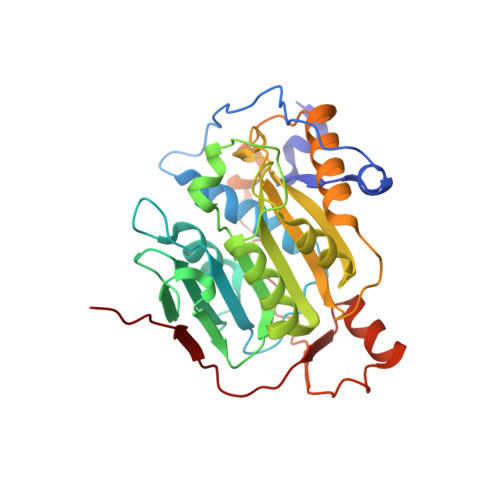 | |
UniProt | |||||
Find proteins for P0DTD1 (Severe acute respiratory syndrome coronavirus 2) Explore P0DTD1 Go to UniProtKB: P0DTD1 | |||||
Entity Groups | |||||
| Sequence Clusters | 30% Identity50% Identity70% Identity90% Identity95% Identity100% Identity | ||||
| UniProt Group | P0DTD1 | ||||
Sequence AnnotationsExpand | |||||
| |||||
Find similar proteins by:
(by identity cutoff) | 3D Structure
Entity ID: 2 | |||||
|---|---|---|---|---|---|
| Molecule | Chains | Sequence Length | Organism | Details | Image |
| Non-structural protein 10 | 141 | Severe acute respiratory syndrome coronavirus 2 | Mutation(s): 0 Gene Names: rep, 1a-1b | 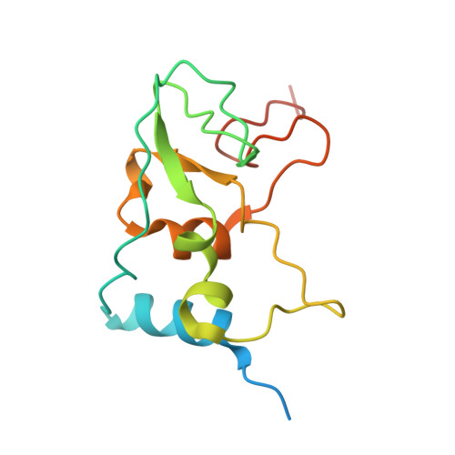 | |
UniProt | |||||
Find proteins for P0DTD1 (Severe acute respiratory syndrome coronavirus 2) Explore P0DTD1 Go to UniProtKB: P0DTD1 | |||||
Entity Groups | |||||
| Sequence Clusters | 30% Identity50% Identity70% Identity90% Identity95% Identity100% Identity | ||||
| UniProt Group | P0DTD1 | ||||
Sequence AnnotationsExpand | |||||
| |||||
Find similar nucleic acids by: Sequence | 3D Structure
Entity ID: 3 | |||||
|---|---|---|---|---|---|
| Molecule | Chains | Length | Organism | Image | |
| RNA (5'-D(*(M7G))-R(P*(A2M)P*UP*UP*A)-3') | 7 | synthetic construct | 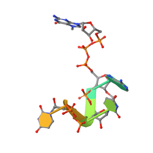 | ||
Sequence AnnotationsExpand | |||||
| |||||
Small Molecules
| Ligands 7 Unique | |||||
|---|---|---|---|---|---|
| ID | Chains | Name / Formula / InChI Key | 2D Diagram | 3D Interactions | |
| SAH (Subject of Investigation/LOI) Query on SAH | G [auth A] | S-ADENOSYL-L-HOMOCYSTEINE C14 H20 N6 O5 S ZJUKTBDSGOFHSH-WFMPWKQPSA-N |  | ||
| BDF Query on BDF | L [auth B] | beta-D-fructopyranose C6 H12 O6 LKDRXBCSQODPBY-ARQDHWQXSA-N |  | ||
| GLC Query on GLC | H [auth A], I [auth A] | alpha-D-glucopyranose C6 H12 O6 WQZGKKKJIJFFOK-DVKNGEFBSA-N |  | ||
| ZN (Subject of Investigation/LOI) Query on ZN | J [auth B], K [auth B] | ZINC ION Zn PTFCDOFLOPIGGS-UHFFFAOYSA-N |  | ||
| FMT Query on FMT | F [auth A] | FORMIC ACID C H2 O2 BDAGIHXWWSANSR-UHFFFAOYSA-N |  | ||
| CL Query on CL | E [auth A] | CHLORIDE ION Cl VEXZGXHMUGYJMC-UHFFFAOYSA-M |  | ||
| MG (Subject of Investigation/LOI) Query on MG | D [auth A], M [auth C] | MAGNESIUM ION Mg JLVVSXFLKOJNIY-UHFFFAOYSA-N |  | ||
Experimental Data & Validation
Experimental Data
- Method: X-RAY DIFFRACTION
- Resolution: 1.78 Å
- R-Value Free: 0.162
- R-Value Work: 0.141
- R-Value Observed: 0.142
- Space Group: P 31 2 1
Unit Cell:
| Length ( Å ) | Angle ( ˚ ) |
|---|---|
| a = 169.351 | α = 90 |
| b = 169.351 | β = 90 |
| c = 52.629 | γ = 120 |
| Software Name | Purpose |
|---|---|
| REFMAC | refinement |
| PDB_EXTRACT | data extraction |
| HKL-3000 | data reduction |
| HKL-3000 | data scaling |
| PHASER | phasing |
Entry History
Deposition Data
- Released Date: 2021-01-06 Deposition Author(s): Minasov, G., Shuvalova, L., Rosas-Lemus, M., Kiryukhina, O., Brunzelle, J.S., Satchell, K.J.F., Center for Structural Genomics of Infectious Diseases (CSGID)
Revision History (Full details and data files)
- Version 1.0: 2021-01-06
Type: Initial release - Version 1.1: 2021-12-15
Changes: Database references - Version 1.2: 2023-10-18
Changes: Data collection, Refinement description - Version 2.0: 2024-03-06
Changes: Data collection, Non-polymer description, Structure summary
