7ADM

Structure of the mycoplasma MIB protein


Experimental Data Snapshot

  • Method: ELECTRON MICROSCOPY
  • Resolution: 3.50 Å
  • Aggregation State: PARTICLE 
  • Reconstruction Method: SINGLE PARTICLE 

wwPDB Validation   3D Report Full Report


This is version 1.0 of the entry. See complete history


Literature

The mycoplasma surface proteins MIB and MIP promote the dissociation of the antibody-antigen interaction.

Nottelet, P.Bataille, L.Gourgues, G.Anger, R.Lartigue, C.Sirand-Pugnet, P.Marza, E.Fronzes, R.Arfi, Y.

(2021) Sci Adv 7

  • DOI: https://doi.org/10.1126/sciadv.abf2403
  • Primary Citation of Related Structures:  
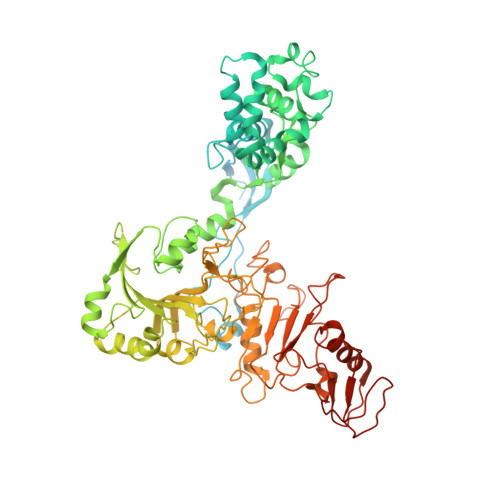
    7ADJ, 7ADK, 7ADM

  • PubMed Abstract: 

    Mycoplasma immunoglobulin binding (MIB) and mycoplasma immunoglobulin protease (MIP) are surface proteins found in the majority of mycoplasma species, acting sequentially to capture antibodies and cleave off their V H domains. Cryo-electron microscopy structures show how MIB and MIP bind to a Fab fragment in a "hug of death" mechanism. As a result, the orientation of the V L and V H domains is twisted out of alignment, disrupting the antigen binding site. We also show that MIB-MIP has the ability to promote the dissociation of the antibody-antigen complex. This system is functional in cells and protects mycoplasmas from antibody-mediated agglutination. These results highlight the key role of the MIB-MIP system in immunity evasion by mycoplasmas through an unprecedented mechanism, and open exciting perspectives to use these proteins as potential tools in the antibody field.


  • Organizational Affiliation

    Structure and Function of Bacterial Nanomachines, UMR 5234, Univ. Bordeaux, CNRS, Institut Européen de Chimie et Biologie, F-33600 Pessac, France.


Macromolecules
Find similar proteins by:  (by identity cutoff)  |  3D Structure
Entity ID: 1
MoleculeChains Sequence LengthOrganismDetailsImage
Putative immunoglobulin-blocking virulence proteinA [auth C]750Mycoplasma mycoides subsp. capriMutation(s): 0 
Gene Names: MMC68F_00609
UniProt
Find proteins for A0A7Z7PMS6 (Mycoplasma mycoides subsp. capri)
Explore A0A7Z7PMS6 
Go to UniProtKB:  A0A7Z7PMS6
Entity Groups  
Sequence Clusters30% Identity50% Identity70% Identity90% Identity95% Identity100% Identity
UniProt GroupA0A7Z7PMS6
Sequence Annotations
Expand
  • Reference Sequence
Experimental Data & Validation

Experimental Data

  • Method: ELECTRON MICROSCOPY
  • Resolution: 3.50 Å
  • Aggregation State: PARTICLE 
  • Reconstruction Method: SINGLE PARTICLE 
EM Software:
TaskSoftware PackageVersion
MODEL REFINEMENTPHENIX
RECONSTRUCTIONcryoSPARC

Structure Validation

View Full Validation Report


Entry History & Funding Information

Deposition Data


Funding OrganizationLocationGrant Number
French National Research AgencyFrance--

Revision History  (Full details and data files)

  • Version 1.0: 2021-04-07
    Type: Initial release