6WF3

Crystal structure of human Naa50 in complex with a cofactor derived inhibitor (compound 1)


Experimental Data Snapshot

  • Method: X-RAY DIFFRACTION
  • Resolution: 2.29 Å
  • R-Value Free: 0.243 
  • R-Value Work: 0.208 
  • R-Value Observed: 0.210 

wwPDB Validation   3D Report Full Report


Ligand Structure Quality Assessment 


This is version 1.1 of the entry. See complete history


Literature

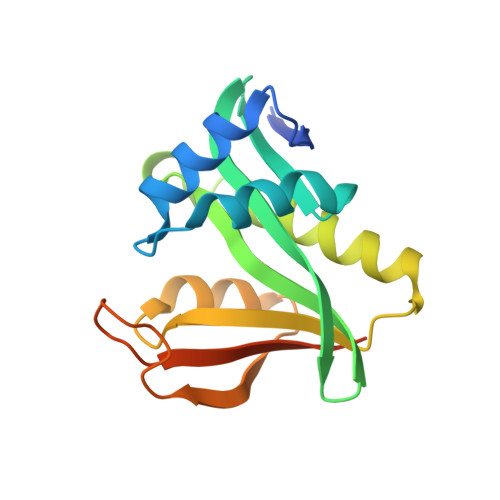
Characterization of SpecificN-alpha-Acetyltransferase 50 (Naa50) Inhibitors Identified Using a DNA Encoded Library.

Kung, P.P.Bingham, P.Burke, B.J.Chen, Q.Cheng, X.Deng, Y.L.Dou, D.Feng, J.Gallego, G.M.Gehring, M.R.Grant, S.K.Greasley, S.Harris, A.R.Maegley, K.A.Meier, J.Meng, X.Montano, J.L.Morgan, B.A.Naughton, B.S.Palde, P.B.Paul, T.A.Richardson, P.Sakata, S.Shaginian, A.Sonnenburg, W.K.Subramanyam, C.Timofeevski, S.Wan, J.Yan, W.Stewart, A.E.

(2020) ACS Med Chem Lett 11: 1175-1184

  • DOI: https://doi.org/10.1021/acsmedchemlett.0c00029
  • Primary Citation of Related Structures:  
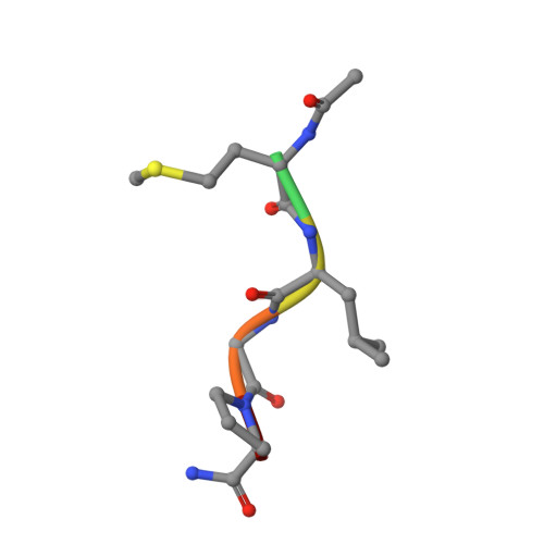
    6WF3, 6WF5, 6WFG, 6WFK, 6WFN, 6WFO

  • PubMed Abstract: 

    Two novel compounds were identified as Naa50 binders/inhibitors using DNA-encoded technology screening. Biophysical and biochemical data as well as cocrystal structures were obtained for both compounds ( 3a and 4a ) to understand their mechanism of action. These data were also used to rationalize the binding affinity differences observed between the two compounds and a MLGP peptide-containing substrate. Cellular target engagement experiments further confirm the Naa50 binding of 4a and demonstrate its selectivity toward related enzymes (Naa10 and Naa60). Additional analogs of inhibitor 4a were also evaluated to study the binding mode observed in the cocrystal structures.


  • Organizational Affiliation

    Worldwide Research and Development, Pfizer Inc., 10770 Science Center Drive, San Diego, California 92121, United States.


Macromolecules
Find similar proteins by:  (by identity cutoff)  |  3D Structure
Entity ID: 1
MoleculeChains Sequence LengthOrganismDetailsImage
N-alpha-acetyltransferase 50
A, B, C, D, E
A, B, C, D, E, F
171Homo sapiensMutation(s): 0 
Gene Names: NAA50MAK3NAT13NAT5
EC: 2.3.1.258 (PDB Primary Data), 2.3.1 (PDB Primary Data)
UniProt & NIH Common Fund Data Resources
Find proteins for Q9GZZ1 (Homo sapiens)
Explore Q9GZZ1 
Go to UniProtKB:  Q9GZZ1
PHAROS:  Q9GZZ1
GTEx:  ENSG00000121579 
Entity Groups  
Sequence Clusters30% Identity50% Identity70% Identity90% Identity95% Identity100% Identity
UniProt GroupQ9GZZ1
Sequence Annotations
Expand
  • Reference Sequence

Find similar proteins by:  Sequence   |   3D Structure  

Entity ID: 2
MoleculeChains Sequence LengthOrganismDetailsImage
ACE-MET-LEU-GLY-PRO-NH2
G, H, I, J, K
G, H, I, J, K, L
6synthetic constructMutation(s): 0 
Sequence Annotations
Expand
  • Reference Sequence
Experimental Data & Validation

Experimental Data

  • Method: X-RAY DIFFRACTION
  • Resolution: 2.29 Å
  • R-Value Free: 0.243 
  • R-Value Work: 0.208 
  • R-Value Observed: 0.210 
  • Space Group: P 1 21 1
Unit Cell:
Length ( Å )Angle ( ˚ )
a = 48.547α = 90
b = 124.17β = 100.92
c = 109.085γ = 90
Software Package:
Software NamePurpose
BUSTERrefinement
XDSdata reduction
autoPROCdata scaling
MOLREPphasing

Structure Validation

View Full Validation Report


Ligand Structure Quality Assessment 


Entry History 

Deposition Data

Revision History  (Full details and data files)

  • Version 1.0: 2020-07-01
    Type: Initial release
  • Version 1.1: 2023-10-18
    Changes: Data collection, Database references, Refinement description