6TQL

Cryo-EM of elastase-treated human uromodulin (UMOD)/Tamm-Horsfall protein (THP) filament


Experimental Data Snapshot

  • Method: ELECTRON MICROSCOPY
  • Resolution: 3.96 Å
  • Aggregation State: FILAMENT 
  • Reconstruction Method: HELICAL 

wwPDB Validation   3D Report Full Report


This is version 1.3 of the entry. See complete history


Literature

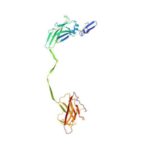
Cryo-EM structure of native human uromodulin, a zona pellucida module polymer.

Stsiapanava, A.Xu, C.Brunati, M.Zamora-Caballero, S.Schaeffer, C.Bokhove, M.Han, L.Hebert, H.Carroni, M.Yasumasu, S.Rampoldi, L.Wu, B.Jovine, L.

(2020) EMBO J 39: e106807-e106807

  • DOI: https://doi.org/10.15252/embj.2020106807
  • Primary Citation of Related Structures:  
    6TQK, 6TQL

  • PubMed Abstract: 

    Assembly of extracellular filaments and matrices mediating fundamental biological processes such as morphogenesis, hearing, fertilization, and antibacterial defense is driven by a ubiquitous polymerization module known as zona pellucida (ZP) "domain". Despite the conservation of this element from hydra to humans, no detailed information is available on the filamentous conformation of any ZP module protein. Here, we report a cryo-electron microscopy study of uromodulin (UMOD)/Tamm-Horsfall protein, the most abundant protein in human urine and an archetypal ZP module-containing molecule, in its mature homopolymeric state. UMOD forms a one-start helix with an unprecedented 180-degree twist between subunits enfolded by interdomain linkers that have completely reorganized as a result of propeptide dissociation. Lateral interaction between filaments in the urine generates sheets exposing a checkerboard of binding sites to capture uropathogenic bacteria, and UMOD-based models of heteromeric vertebrate egg coat filaments identify a common sperm-binding region at the interface between subunits.


  • Organizational Affiliation

    Department of Biosciences and Nutrition, Karolinska Institutet, Huddinge, Sweden.


Macromolecules
Find similar proteins by:  (by identity cutoff)  |  3D Structure
Entity ID: 1
MoleculeChains Sequence LengthOrganismDetailsImage
Uromodulin
A, B, C
296Homo sapiensMutation(s): 0 
UniProt & NIH Common Fund Data Resources
Find proteins for P07911 (Homo sapiens)
Explore P07911 
Go to UniProtKB:  P07911
PHAROS:  P07911
GTEx:  ENSG00000169344 
Entity Groups  
Sequence Clusters30% Identity50% Identity70% Identity90% Identity95% Identity100% Identity
UniProt GroupP07911
Sequence Annotations
Expand
  • Reference Sequence
Oligosaccharides

Help

Entity ID: 2
MoleculeChains Length2D Diagram Glycosylation3D Interactions
2-acetamido-2-deoxy-beta-D-glucopyranose-(1-4)-2-acetamido-2-deoxy-beta-D-glucopyranose
D, F
2N-Glycosylation
Glycosylation Resources
GlyTouCan:  G42666HT
GlyCosmos:  G42666HT
GlyGen:  G42666HT
Entity ID: 3
MoleculeChains Length2D Diagram Glycosylation3D Interactions
beta-D-galactopyranose-(1-4)-2-acetamido-2-deoxy-beta-D-glucopyranose-(1-2)-[2-acetamido-2-deoxy-beta-D-glucopyranose-(1-4)]alpha-D-mannopyranose-(1-6)-[2-acetamido-2-deoxy-beta-D-glucopyranose-(1-2)-[2-acetamido-2-deoxy-beta-D-glucopyranose-(1-4)]alpha-D-mannopyranose-(1-3)]beta-D-mannopyranose-(1-4)-2-acetamido-2-deoxy-beta-D-glucopyranose-(1-4)-2-acetamido-2-deoxy-beta-D-glucopyranose
E
10N-Glycosylation
Glycosylation Resources
GlyTouCan:  G88219FI
GlyCosmos:  G88219FI
GlyGen:  G88219FI
Entity ID: 4
MoleculeChains Length2D Diagram Glycosylation3D Interactions
2-acetamido-2-deoxy-beta-D-glucopyranose-(1-2)-[2-acetamido-2-deoxy-beta-D-glucopyranose-(1-4)]alpha-D-mannopyranose-(1-3)-[2-acetamido-2-deoxy-beta-D-glucopyranose-(1-4)-alpha-D-mannopyranose-(1-6)]beta-D-mannopyranose-(1-4)-2-acetamido-2-deoxy-beta-D-glucopyranose-(1-4)-2-acetamido-2-deoxy-beta-D-glucopyranose
G
8N-Glycosylation
Glycosylation Resources
GlyTouCan:  G64652EG
GlyCosmos:  G64652EG
GlyGen:  G64652EG
Small Molecules
Ligands 1 Unique
IDChains Name / Formula / InChI Key2D Diagram3D Interactions
NAG
Query on NAG

Download Ideal Coordinates CCD File 
H [auth A],
I [auth B]
2-acetamido-2-deoxy-beta-D-glucopyranose
C8 H15 N O6
OVRNDRQMDRJTHS-FMDGEEDCSA-N
Experimental Data & Validation

Experimental Data

  • Method: ELECTRON MICROSCOPY
  • Resolution: 3.96 Å
  • Aggregation State: FILAMENT 
  • Reconstruction Method: HELICAL 
EM Software:
TaskSoftware PackageVersion
MODEL REFINEMENTPHENIX
RECONSTRUCTIONRELION3.0.5
RECONSTRUCTIONPHENIX

Structure Validation

View Full Validation Report


Entry History & Funding Information

Deposition Data


Funding OrganizationLocationGrant Number
Swedish Research CouncilSweden2016-03999
Knut and Alice Wallenberg FoundationSweden2018.0042
Ministry of Education (MoE, Singapore)Singapore2017-T1-001-168
Ministry of Education (MoE, Singapore)Singapore2016-T2-1-010

Revision History  (Full details and data files)

  • Version 1.0: 2020-11-04
    Type: Initial release
  • Version 1.1: 2020-11-25
    Changes: Database references
  • Version 1.2: 2020-12-16
    Changes: Database references
  • Version 1.3: 2021-03-03
    Changes: Database references