


Chimera of bacteriophage OBP gp146 central spike protein and a T4 gp5 beta-helix fragment
Buth, S.A., Shneider, M.M., Leiman, P.G.To be published.
Experimental Data Snapshot
Entity ID: 1 | |||||
|---|---|---|---|---|---|
| Molecule | Chains | Sequence Length | Organism | Details | Image |
| CHIMERA OF BACTERIOPHAGE OBP GP146 AND A T4 GP5 | 148 | Tequatrovirus T4, Pseudomonas phage OBP This entity is chimeric | Mutation(s): 0 EC: 3.2.1.17 | 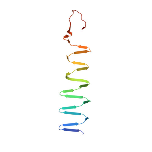 | |
UniProt | |||||
Find proteins for G9IA38 (Pseudomonas phage OBP) Explore G9IA38 Go to UniProtKB: G9IA38 | |||||
Find proteins for P16009 (Enterobacteria phage T4) Explore P16009 Go to UniProtKB: P16009 | |||||
Entity Groups | |||||
| Sequence Clusters | 30% Identity50% Identity70% Identity90% Identity95% Identity100% Identity | ||||
| UniProt Groups | G9IA38P16009 | ||||
Sequence AnnotationsExpand | |||||
| |||||
| Ligands 5 Unique | |||||
|---|---|---|---|---|---|
| ID | Chains | Name / Formula / InChI Key | 2D Diagram | 3D Interactions | |
| STE Query on STE | H [auth A], N [auth D] | STEARIC ACID C18 H36 O2 QIQXTHQIDYTFRH-UHFFFAOYSA-N |  | ||
| ELA Query on ELA | I [auth A], O [auth F] | 9-OCTADECENOIC ACID C18 H34 O2 ZQPPMHVWECSIRJ-MDZDMXLPSA-N |  | ||
| PLM Query on PLM | J [auth B], P [auth F] | PALMITIC ACID C16 H32 O2 IPCSVZSSVZVIGE-UHFFFAOYSA-N |  | ||
| FE2 Query on FE2 | G [auth A], L [auth D] | FE (II) ION Fe CWYNVVGOOAEACU-UHFFFAOYSA-N |  | ||
| MG Query on MG | K [auth C], M [auth D] | MAGNESIUM ION Mg JLVVSXFLKOJNIY-UHFFFAOYSA-N |  | ||
| Length ( Å ) | Angle ( ˚ ) |
|---|---|
| a = 140.013 | α = 90 |
| b = 140.013 | β = 90 |
| c = 63.161 | γ = 90 |
| Software Name | Purpose |
|---|---|
| PHENIX | refinement |
| XDS | data reduction |
| XDS | data scaling |
| PHASER | phasing |
| Funding Organization | Location | Grant Number |
|---|---|---|
| Swiss National Science Foundation | Switzerland | 310030_144243 |
RCSB PDB (citation) is hosted by
RCSB PDB is a member of the

