6NOH

Crystal structure of FBF-2 repeat 5 mutant (C363S, R364Y, Q367S) in complex with 8-nt RNA


Experimental Data Snapshot

  • Method: X-RAY DIFFRACTION
  • Resolution: 2.25 Å
  • R-Value Free: 0.206 
  • R-Value Work: 0.159 
  • R-Value Observed: 0.163 

wwPDB Validation   3D Report Full Report

Currently 6NOH does not have a validation slider image.


This is version 1.2 of the entry. See complete history


Literature

Engineering a conserved RNA regulatory protein repurposes its biological function in vivo .

Bhat, V.D.McCann, K.L.Wang, Y.Fonseca, D.R.Shukla, T.Alexander, J.C.Qiu, C.Wickens, M.Lo, T.W.Tanaka Hall, T.M.Campbell, Z.T.

(2019) Elife 8

  • DOI: https://doi.org/10.7554/eLife.43788
  • Primary Citation of Related Structures:  
    6NOC, 6NOD, 6NOF, 6NOH

  • PubMed Abstract: 

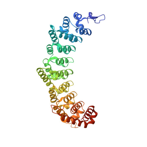
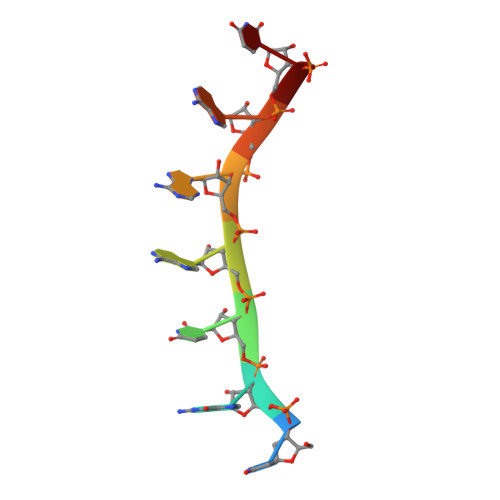
    PUF ( PU milio/ F BF) RNA-binding proteins recognize distinct elements. In C. elegans , PUF-8 binds to an 8-nt motif and restricts proliferation in the germline. Conversely, FBF-2 recognizes a 9-nt element and promotes mitosis. To understand how motif divergence relates to biological function, we first determined a crystal structure of PUF-8. Comparison of this structure to that of FBF-2 revealed a major difference in a central repeat. We devised a modified yeast 3-hybrid screen to identify mutations that confer recognition of an 8-nt element to FBF-2. We identified several such mutants and validated structurally and biochemically their binding to 8-nt RNA elements. Using genome engineering, we generated a mutant animal with a substitution in FBF-2 that confers preferential binding to the PUF-8 element. The mutant largely rescued overproliferation in animals that spontaneously generate tumors in the absence of puf-8 . This work highlights the critical role of motif length in the specification of biological function.


  • Organizational Affiliation

    Department of Biological Sciences, University of Texas Dallas, Richardson, United States.


Macromolecules

Find similar proteins by:  (by identity cutoff)  |  3D Structure
Entity ID: 1
MoleculeChains Sequence LengthOrganismDetailsImage
Fem-3 mRNA-binding factor 2413Caenorhabditis elegansMutation(s): 3 
Gene Names: fbf-2F21H12.5
UniProt
Find proteins for Q09312 (Caenorhabditis elegans)
Explore Q09312 
Go to UniProtKB:  Q09312
Entity Groups  
Sequence Clusters30% Identity50% Identity70% Identity90% Identity95% Identity100% Identity
UniProt GroupQ09312
Sequence Annotations
Expand
  • Reference Sequence

Find similar nucleic acids by:  Sequence   |   3D Structure  

Entity ID: 2
MoleculeChains LengthOrganismImage
RNA (5'-R(*UP*GP*UP*AP*AP*AP*UP*A)-3')8Caenorhabditis elegans
Sequence Annotations
Expand
  • Reference Sequence
Small Molecules
Ligands 1 Unique
IDChains Name / Formula / InChI Key2D Diagram3D Interactions
EDO
Query on EDO

Download Ideal Coordinates CCD File 
C [auth A]
D [auth A]
E [auth A]
F [auth A]
G [auth A]
C [auth A],
D [auth A],
E [auth A],
F [auth A],
G [auth A],
H [auth A],
I [auth A],
J [auth A],
K [auth A],
L [auth A],
M [auth A],
N [auth A],
O [auth B]
1,2-ETHANEDIOL
C2 H6 O2
LYCAIKOWRPUZTN-UHFFFAOYSA-N
Experimental Data & Validation

Experimental Data

  • Method: X-RAY DIFFRACTION
  • Resolution: 2.25 Å
  • R-Value Free: 0.206 
  • R-Value Work: 0.159 
  • R-Value Observed: 0.163 
  • Space Group: P 61
Unit Cell:
Length ( Å )Angle ( ˚ )
a = 96.397α = 90
b = 96.397β = 90
c = 99.853γ = 120
Software Package:
Software NamePurpose
PHENIXrefinement
SCALEPACKdata scaling
PDB_EXTRACTdata extraction
HKL-2000data reduction
PHASERphasing

Structure Validation

View Full Validation Report

Currently 6NOH does not have a validation slider image.


Entry History 

Deposition Data

Revision History  (Full details and data files)

  • Version 1.0: 2019-01-30
    Type: Initial release
  • Version 1.1: 2020-10-07
    Changes: Database references
  • Version 1.2: 2023-10-11
    Changes: Data collection, Database references, Refinement description