6MQW

Crystal Structure of All-trans Retinal-Bound R111K:Y134F:T54V:R132Q:P39Y:R59Y:L121E Human Cellular Retinoic Acid Binding Protein II Irradiated with 400 nm laser (30 seconds) and subsequently dark adapted (10 minutes) at 2.1 Angstrom Resolution


Experimental Data Snapshot

  • Method: X-RAY DIFFRACTION
  • Resolution: 2.09 Å
  • R-Value Free: 0.220 
  • R-Value Work: 0.183 
  • R-Value Observed: 0.186 

wwPDB Validation   3D Report Full Report

Currently 6MQW does not have a validation slider image.


This is version 1.3 of the entry. See complete history


Literature

Mimicking Microbial Rhodopsin Isomerization in a Single Crystal.

Ghanbarpour, A.Nairat, M.Nosrati, M.Santos, E.M.Vasileiou, C.Dantus, M.Borhan, B.Geiger, J.H.

(2019) J Am Chem Soc 141: 1735-1741

  • DOI: https://doi.org/10.1021/jacs.8b12493
  • Primary Citation of Related Structures:  
    6MOP, 6MOQ, 6MOR, 6MOV, 6MOX, 6MPK, 6MQI, 6MQJ, 6MQW, 6MQX, 6MQY, 6MQZ, 6MR0

  • PubMed Abstract: 

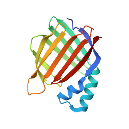
    Bacteriorhodopsin represents the simplest, and possibly most abundant, phototropic system requiring only a retinal-bound transmembrane protein to convert photons of light to an energy-generating proton gradient. The creation and interrogation of a microbial rhodopsin mimic, based on an orthogonal protein system, would illuminate the design elements required to generate new photoactive proteins with novel function. We describe a microbial rhodopsin mimic, created using a small soluble protein as a template, that specifically photoisomerizes all- trans to 13- cis retinal followed by thermal relaxation to the all- trans isomer, mimicking the bacteriorhodopsin photocycle, in a single crystal. The key element for selective isomerization is a tuned steric interaction between the chromophore and protein, similar to that seen in the microbial rhodopsins. It is further demonstrated that a single mutation converts the system to a protein photoswitch without chromophore photoisomerization or conformational change.


  • Organizational Affiliation

    Michigan State University , Department of Chemistry , East Lansing , Michigan 48824 , United States.


Macromolecules
Find similar proteins by:  (by identity cutoff)  |  3D Structure
Entity ID: 1
MoleculeChains Sequence LengthOrganismDetailsImage
Cellular retinoic acid-binding protein 2137Homo sapiensMutation(s): 7 
Gene Names: CRABP2
UniProt & NIH Common Fund Data Resources
Find proteins for P29373 (Homo sapiens)
Explore P29373 
Go to UniProtKB:  P29373
PHAROS:  P29373
GTEx:  ENSG00000143320 
Entity Groups  
Sequence Clusters30% Identity50% Identity70% Identity90% Identity95% Identity100% Identity
UniProt GroupP29373
Sequence Annotations
Expand
  • Reference Sequence
Small Molecules
Ligands 1 Unique
IDChains Name / Formula / InChI Key2D Diagram3D Interactions
RET
Query on RET

Download Ideal Coordinates CCD File 
B [auth A]RETINAL
C20 H28 O
NCYCYZXNIZJOKI-OVSJKPMPSA-N
Experimental Data & Validation

Experimental Data

  • Method: X-RAY DIFFRACTION
  • Resolution: 2.09 Å
  • R-Value Free: 0.220 
  • R-Value Work: 0.183 
  • R-Value Observed: 0.186 
  • Space Group: P 31 2 1
Unit Cell:
Length ( Å )Angle ( ˚ )
a = 58.586α = 90
b = 58.586β = 90
c = 100.178γ = 120
Software Package:
Software NamePurpose
PHENIXrefinement
HKL-2000data reduction
HKL-2000data scaling
PHASERphasing

Structure Validation

View Full Validation Report

Currently 6MQW does not have a validation slider image.


Entry History & Funding Information

Deposition Data


Funding OrganizationLocationGrant Number
National Institutes of Health/National Human Genome Research Institute (NIH/NHGRI)United States--

Revision History  (Full details and data files)

  • Version 1.0: 2019-01-09
    Type: Initial release
  • Version 1.1: 2019-02-13
    Changes: Data collection, Database references
  • Version 1.2: 2019-12-18
    Changes: Author supporting evidence
  • Version 1.3: 2023-10-11
    Changes: Data collection, Database references, Refinement description