A DNA-Binding Protein Tunes Septum Placement during Bacillus subtilis Sporulation.
Brown, E.E., Miller, A.K., Krieger, I.V., Otto, R.M., Sacchettini, J.C., Herman, J.K.(2019) J Bacteriol 201
- PubMed: 31160399
- DOI: https://doi.org/10.1128/JB.00287-19
- Primary Citation of Related Structures:
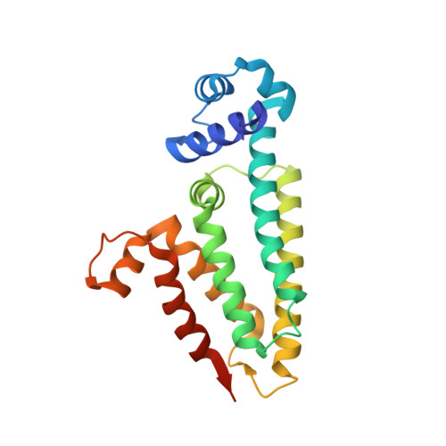
6MJ1 - PubMed Abstract:
Bacillus subtilis is a bacterium capable of differentiating into a spore form more resistant to environmental stress. Early in sporulation, each cell possesses two copies of a circular chromosome. A polar FtsZ ring (Z ring) directs septation over one of the chromosomes, generating two cell compartments. The smaller "forespore" compartment initially contains only 25 to 30% of one chromosome, and this transient genetic asymmetry is required for differentiation. Timely assembly of polar Z rings and precise capture of the chromosome in the forespore both require the DNA-binding protein RefZ. To mediate its role in chromosome capture, RefZ must bind to specific DNA motifs ( RBM s) that localize near the poles at the time of septation. Cells artificially induced to express RefZ during vegetative growth cannot assemble Z rings, an effect that also requires DNA binding. We hypothesized that RefZ- RBM complexes mediate precise chromosome capture by modulating FtsZ function. To investigate, we isolated 10 RefZ loss-of-function (rLOF) variants unable to inhibit cell division yet still capable of binding RBM s. Sporulating cells expressing the rLOF variants in place of wild-type RefZ phenocopied a Δ refZ mutant, suggesting that RefZ acts through an FtsZ-dependent mechanism. The crystal structure of RefZ was solved, and wild-type RefZ and the rLOF variants were further characterized. Our data suggest that RefZ's oligomerization state and specificity for the RBM s are critical determinants influencing RefZ's ability to affect FtsZ dynamics. We propose that RBM -bound RefZ complexes function as a developmentally regulated nucleoid occlusion system for fine-tuning the position of the septum relative to the chromosome during sporulation. IMPORTANCE The bacterial nucleoid forms a large, highly organized structure. Thus, in addition to storing the genetic code, the nucleoid harbors positional information that can be leveraged by DNA-binding proteins to spatially constrain cellular activities. During B. subtilis sporulation, the nucleoid undergoes reorganization, and the cell division protein FtsZ assembles polarly to direct septation over one chromosome. The TetR family protein RefZ binds DNA motifs ( RBM s) localized near the poles at the time of division and is required for both timely FtsZ assembly and precise capture of DNA in the future spore compartment. Our data suggest that RefZ exploits nucleoid organization by associating with polarly localized RBM s to modulate the positioning of FtsZ relative to the chromosome during sporulation.
Organizational Affiliation:
Department of Biochemistry and Biophysics, Texas A&M University, College Station, Texas, USA.