6LL6

Crsyal structure of EcFtsZ (residues 11-316)


Experimental Data Snapshot

  • Method: X-RAY DIFFRACTION
  • Resolution: 2.50 Å
  • R-Value Free: 0.241 
  • R-Value Work: 0.184 
  • R-Value Observed: 0.186 

wwPDB Validation   3D Report Full Report


Ligand Structure Quality Assessment 


This is version 1.2 of the entry. See complete history


Literature

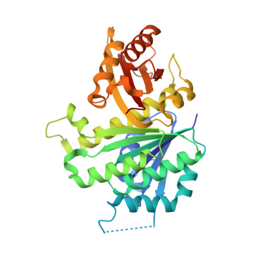
Crystal structures of the cell-division protein FtsZ from Klebsiella pneumoniae and Escherichia coli.

Yoshizawa, T.Fujita, J.Terakado, H.Ozawa, M.Kuroda, N.Tanaka, S.I.Uehara, R.Matsumura, H.

(2020) Acta Crystallogr F Struct Biol Commun 76: 86-93

  • DOI: https://doi.org/10.1107/S2053230X2000076X
  • Primary Citation of Related Structures:  
    6LL5, 6LL6

  • PubMed Abstract: 

    FtsZ, a tubulin-like GTPase, is essential for bacterial cell division. In the presence of GTP, FtsZ polymerizes into filamentous structures, which are key to generating force in cell division. However, the structural basis for the molecular mechanism underlying FtsZ function remains to be elucidated. In this study, crystal structures of the enzymatic domains of FtsZ from Klebsiella pneumoniae (KpFtsZ) and Escherichia coli (EcFtsZ) were determined at 1.75 and 2.50 Å resolution, respectively. Both FtsZs form straight protofilaments in the crystals, and the two structures adopted relaxed (R) conformations. The T3 loop, which is involved in GTP/GDP binding and FtsZ assembly/disassembly, adopted a unique open conformation in KpFtsZ, while the T3 loop of EcFtsZ was partially disordered. The crystal structure of EcFtsZ can explain the results from previous functional analyses using EcFtsZ mutants.


  • Organizational Affiliation

    Department of Biotechnology, College of Life Sciences, Ritsumeikan University, 1-1-1 Noji-higashi, Kusatsu, Shiga 525-8577, Japan.


Macromolecules
Find similar proteins by:  (by identity cutoff)  |  3D Structure
Entity ID: 1
MoleculeChains Sequence LengthOrganismDetailsImage
Cell division protein FtsZ309Escherichia coliMutation(s): 0 
Gene Names: ftsZDNQ45_06620
UniProt
Find proteins for P0A9A6 (Escherichia coli (strain K12))
Explore P0A9A6 
Go to UniProtKB:  P0A9A6
Entity Groups  
Sequence Clusters30% Identity50% Identity70% Identity90% Identity95% Identity100% Identity
UniProt GroupP0A9A6
Sequence Annotations
Expand
  • Reference Sequence
Small Molecules
Ligands 1 Unique
IDChains Name / Formula / InChI Key2D Diagram3D Interactions
GDP (Subject of Investigation/LOI)
Query on GDP

Download Ideal Coordinates CCD File 
B [auth A]GUANOSINE-5'-DIPHOSPHATE
C10 H15 N5 O11 P2
QGWNDRXFNXRZMB-UUOKFMHZSA-N
Experimental Data & Validation

Experimental Data

  • Method: X-RAY DIFFRACTION
  • Resolution: 2.50 Å
  • R-Value Free: 0.241 
  • R-Value Work: 0.184 
  • R-Value Observed: 0.186 
  • Space Group: P 1
Unit Cell:
Length ( Å )Angle ( ˚ )
a = 38.28α = 88.789
b = 41.67β = 72.057
c = 45.03γ = 72.445
Software Package:
Software NamePurpose
PHENIXrefinement
XDSdata reduction
XDSdata scaling
PHASERphasing

Structure Validation

View Full Validation Report


Ligand Structure Quality Assessment 


Entry History & Funding Information

Deposition Data


Funding OrganizationLocationGrant Number
Japan Society for the Promotion of Science (JSPS)Japan15J00589
Japan Agency for Medical Research and Development (AMED)Japan19am0101070

Revision History  (Full details and data files)

  • Version 1.0: 2020-02-26
    Type: Initial release
  • Version 1.1: 2024-03-27
    Changes: Data collection, Database references
  • Version 1.2: 2024-04-03
    Changes: Refinement description