6IIU

Crystal structure of the human thromboxane A2 receptor bound to ramatroban


Experimental Data Snapshot

  • Method: X-RAY DIFFRACTION
  • Resolution: 2.50 Å
  • R-Value Free: 0.218 
  • R-Value Work: 0.198 
  • R-Value Observed: 0.199 

wwPDB Validation   3D Report Full Report


Ligand Structure Quality Assessment 


This is version 1.2 of the entry. See complete history


Literature

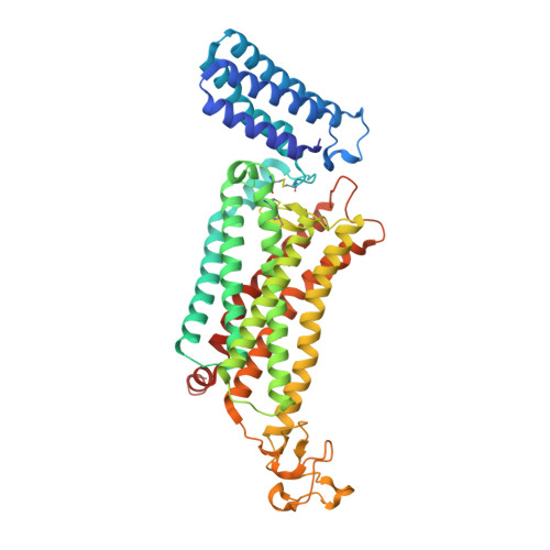
Structural basis for ligand recognition of the human thromboxane A2receptor.

Fan, H.Chen, S.Yuan, X.Han, S.Zhang, H.Xia, W.Xu, Y.Zhao, Q.Wu, B.

(2019) Nat Chem Biol 15: 27-33

  • DOI: https://doi.org/10.1038/s41589-018-0170-9
  • Primary Citation of Related Structures:  
    6IIU, 6IIV

  • PubMed Abstract: 

    Stimulated by thromboxane A 2 , an endogenous arachidonic acid metabolite, the thromboxane A 2 receptor (TP) plays a pivotal role in cardiovascular homeostasis, and thus is considered as an important drug target for cardiovascular disease. Here, we report crystal structures of the human TP bound to two nonprostanoid antagonists, ramatroban and daltroban, at 2.5 Å and 3.0 Å resolution, respectively. The TP structures reveal a ligand-binding pocket capped by two layers of extracellular loops that are stabilized by two disulfide bonds, limiting ligand access from the extracellular milieu. These structures provide details of interactions between the receptor and antagonists, which help to integrate previous mutagenesis and SAR data. Molecular docking of prostanoid-like ligands, combined with mutagenesis, ligand-binding and functional assays, suggests a prostanoid binding mode that may also be adopted by other prostanoid receptors. These insights into TP deepen our understanding about ligand recognition and selectivity mechanisms of this physiologically important receptor.


  • Organizational Affiliation

    CAS Key Laboratory of Receptor Research, Shanghai Institute of Materia Medica, Chinese Academy of Sciences, Shanghai, China.


Macromolecules
Find similar proteins by:  (by identity cutoff)  |  3D Structure
Entity ID: 1
MoleculeChains Sequence LengthOrganismDetailsImage
Soluble cytochrome b562,Thromboxane A2 receptor,Rubredoxin,Thromboxane A2 receptor484Escherichia coliHomo sapiensClostridium pasteurianumMutation(s): 4 
Gene Names: cybCTBXA2R
Membrane Entity: Yes 
UniProt & NIH Common Fund Data Resources
Find proteins for P0ABE7 (Escherichia coli)
Explore P0ABE7 
Go to UniProtKB:  P0ABE7
Find proteins for P00268 (Clostridium pasteurianum)
Explore P00268 
Go to UniProtKB:  P00268
Find proteins for P21731 (Homo sapiens)
Explore P21731 
Go to UniProtKB:  P21731
PHAROS:  P21731
GTEx:  ENSG00000006638 
Entity Groups  
Sequence Clusters30% Identity50% Identity70% Identity90% Identity95% Identity100% Identity
UniProt GroupsP00268P0ABE7P21731
Sequence Annotations
Expand
  • Reference Sequence
Small Molecules
Ligands 5 Unique
IDChains Name / Formula / InChI Key2D Diagram3D Interactions
A8X
Query on A8X

Download Ideal Coordinates CCD File 
B [auth A]3-[(3R)-3-[(4-fluorophenyl)sulfonylamino]-1,2,3,4-tetrahydrocarbazol-9-yl]propanoic acid
C21 H21 F N2 O4 S
LDXDSHIEDAPSSA-OAHLLOKOSA-N
CLR
Query on CLR

Download Ideal Coordinates CCD File 
D [auth A]CHOLESTEROL
C27 H46 O
HVYWMOMLDIMFJA-DPAQBDIFSA-N
OLC
Query on OLC

Download Ideal Coordinates CCD File 
E [auth A],
F [auth A]
(2R)-2,3-dihydroxypropyl (9Z)-octadec-9-enoate
C21 H40 O4
RZRNAYUHWVFMIP-GDCKJWNLSA-N
GOL
Query on GOL

Download Ideal Coordinates CCD File 
G [auth A]GLYCEROL
C3 H8 O3
PEDCQBHIVMGVHV-UHFFFAOYSA-N
ZN
Query on ZN

Download Ideal Coordinates CCD File 
C [auth A]ZINC ION
Zn
PTFCDOFLOPIGGS-UHFFFAOYSA-N
Binding Affinity Annotations 
IDSourceBinding Affinity
A8X Binding MOAD:  6IIU Ki: 11 (nM) from 1 assay(s)
Experimental Data & Validation

Experimental Data

  • Method: X-RAY DIFFRACTION
  • Resolution: 2.50 Å
  • R-Value Free: 0.218 
  • R-Value Work: 0.198 
  • R-Value Observed: 0.199 
  • Space Group: C 2 2 21
Unit Cell:
Length ( Å )Angle ( ˚ )
a = 81.16α = 90
b = 155.08β = 90
c = 128.13γ = 90
Software Package:
Software NamePurpose
BUSTERrefinement
XDSdata reduction
XDSdata scaling
PHASERphasing

Structure Validation

View Full Validation Report


Ligand Structure Quality Assessment 


Entry History & Funding Information

Deposition Data


Funding OrganizationLocationGrant Number
Chinese Academy of SciencesChinaQYZDB-SSW-SMC024

Revision History  (Full details and data files)

  • Version 1.0: 2018-12-19
    Type: Initial release
  • Version 1.1: 2018-12-26
    Changes: Data collection, Database references
  • Version 1.2: 2023-11-22
    Changes: Data collection, Database references, Refinement description