TapA acts as specific chaperone in TasA filament formation by strand complementation.
Roske, Y., Lindemann, F., Diehl, A., Cremer, N., Higman, V.A., Schlegel, B., Leidert, M., Driller, K., Turgay, K., Schmieder, P., Heinemann, U., Oschkinat, H.(2023) Proc Natl Acad Sci U S A 120: e2217070120-e2217070120
- PubMed: 37068239
- DOI: https://doi.org/10.1073/pnas.2217070120
- Primary Citation of Related Structures:
6HQC, 6QAY, 8AIF - PubMed Abstract:
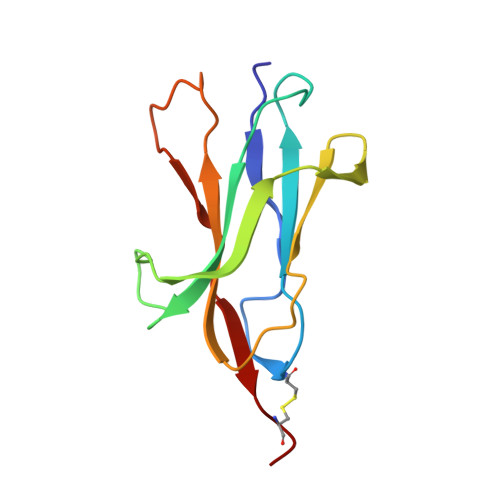
Studying mechanisms of bacterial biofilm generation is of vital importance to understanding bacterial cell-cell communication, multicellular cohabitation principles, and the higher resilience of microorganisms in a biofilm against antibiotics. Biofilms of the nonpathogenic, gram-positive soil bacterium Bacillus subtilis serve as a model system with biotechnological potential toward plant protection. Its major extracellular matrix protein components are TasA and TapA. The nature of TasA filaments has been of debate, and several forms, amyloidic and non-Thioflavin T-stainable have been observed. Here, we present the three-dimensional structure of TapA and uncover the mechanism of TapA-supported growth of nonamyloidic TasA filaments. By analytical ultracentrifugation and NMR, we demonstrate TapA-dependent acceleration of filament formation from solutions of folded TasA. Solid-state NMR revealed intercalation of the N-terminal TasA peptide segment into subsequent protomers to form a filament composed of β-sandwich subunits. The secondary structure around the intercalated N-terminal strand β0 is conserved between filamentous TasA and the Fim and Pap proteins, which form bacterial type I pili, demonstrating such construction principles in a gram-positive organism. Analogous to the chaperones of the chaperone-usher pathway, the role of TapA is in donating its N terminus to serve for TasA folding into an Ig domain-similar filament structure by donor-strand complementation. According to NMR and since the V-set Ig fold of TapA is already complete, its participation within a filament beyond initiation is unlikely. Intriguingly, the most conserved residues in TasA-like proteins (camelysines) of Bacillaceae are located within the protomer interface.
Organizational Affiliation:
Structural Biology, Max Delbrück Center for Molecular Medicine, 13125 Berlin, Germany.