Manipulating the Folding Landscape of a Multidomain Protein.
Kantaev, R., Riven, I., Goldenzweig, A., Barak, Y., Dym, O., Peleg, Y., Albeck, S., Fleishman, S.J., Haran, G.(2018) J Phys Chem B 122: 11030-11038
- PubMed: 30088929
- DOI: https://doi.org/10.1021/acs.jpcb.8b04834
- Primary Citation of Related Structures:
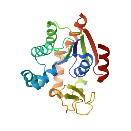
6HAM, 6HAP - PubMed Abstract:
Folding of proteins to their functional conformation is paramount to life. Though 75% of the proteome consists of multidomain proteins, our knowledge of folding has been based primarily on studies conducted on single-domain and fast-folding proteins. Nonetheless, the complexity of folding landscapes exhibited by multidomain proteins has received increased scrutiny in recent years. We study the three-domain protein adenylate kinase from E. coli (AK), which has been shown to fold through a series of pathways involving several intermediate states. We use a protein design method to manipulate the folding landscape of AK, and single-molecule FRET spectroscopy to study the effects on the folding process. Mutations introduced in the NMP binding (NMPbind) domain of the protein are found to have unexpected effects on the folding landscape. Thus, while stabilizing mutations in the core of the NMPbind domain retain the main folding pathways of wild-type AK, a destabilizing mutation at the interface between the NMPbind and the CORE domains causes a significant repartition of the flux between the folding pathways. Our results demonstrate the outstanding plasticity of the folding landscape of AK and reveal how specific mutations in the primary structure are translated into changes in folding dynamics. The combination of methodologies introduced in this work should prove useful for deepening our understanding of the folding process of multidomain proteins.